Notícias

RapAdmin

Skeletal muscle aging, clinically recognized as sarcopenia, has traditionally been viewed through the lens of passive “wear and tear.” However, recent evidence definitively shifts this paradigm toward an active, pathology-driving mechanism: cellular senescence. This review synthesizes mounting data demonstrating that the accumulation of senescent cells across various muscle compartments directly orchestrates the progressive loss of muscle mass, strength, and regenerative capacity.

Crucially, senescence in skeletal muscle is not confined to a single cell type. It actively infiltrates muscle stem cells (MuSCs), fibro-adipogenic progenitors (FAPs), immune cells, and even terminally differentiated, post-mitotic myofibers. Once senescent, these cells exhibit profound chromatin alterations, resistance to apoptosis, and the secretion of a toxic senescence-associated secretory phenotype (SASP). The SASP, a cocktail of pro-inflammatory cytokines, chemokines, and extracellular matrix-modifying enzymes, propagates chronic inflammation (“inflammaging”), disrupts niche homeostasis, and induces secondary senescence in neighboring healthy cells.

For example, senescent FAPs lose the ability to secrete WISP1—a vital regenerative signal—while simultaneously driving fibrotic tissue remodeling via transforming growth factor-β (TGF-β) secretion. Concurrently, senescent MuSCs suffer from impaired autophagy and profound regenerative deficits. Even post-mitotic myofibers, previously thought immune to classic replicative senescence, exhibit stress-induced premature senescence driven by p21CIP1 expression, further exacerbating tissue decline.

This mechanistic clarity has accelerated the development of senotherapeutics as a countermeasure. Senolytics (like the BCL-2/BCL-XL inhibitor ABT263 and CAR-T cells targeting uPAR) selectively induce apoptosis in senescent cells, thereby rejuvenating MuSC function in preclinical models. Senomorphics (such as the CCR5 antagonist Maraviroc and natural polyphenols) aim to suppress the SASP without eliminating the cells. While these pharmacological interventions present a formidable frontier against age-related frailty, clinical translation remains gated by target specificity, the complex heterogeneity of the senescent niche, and the potential off-target consequences of broad senescent cell clearance

Source

Leitura Relacionada: HIV Drug (Maraviroc) Reverses Muscle Aging by purging “Zombie Cell” Signals

RapAdmin

Recent large-scale genetic analyses reveal that lifelong, partial inhibition of myostatin—a well-known negative regulator of muscle growth—significantly increases skeletal muscle mass and strength while reciprocally decreasing body fat. Leveraging exome sequencing and deep phenotyping data from 1.1 million participants across 11 human cohorts, researchers identified 13,454 individuals carrying rare, function-disrupting mutations in the myostatin (MSTN) gene.

The primary finding is highly actionable for the longevity and biotech sectors: heterozygous carriers of predicted loss-of-function (pLoF) and function-disrupting missense variants display a distinct morphological advantage. These individuals possess greater fat-free mass, higher grip strength, and decreased overall adiposity compared to non-carriers. Advanced validation using whole-body MRI on a subset of 77,572 individuals confirmed that these genetic variations result in an average 5–8% increase in overall muscle mass, with specific muscle groups, such as the gluteus, showing volume increases in excess of 10%.

Importantly, this phenotype does not appear to carry the cardiometabolic or reproductive penalties previously hypothesized to accompany myostatin suppression. The data demonstrate no elevated risk for hypertrophic cardiomyopathy, heart failure, or polycystic ovary syndrome (PCOS) among carriers. This structural and functional validation is particularly relevant for modern clinical applications, notably the pressing need to preserve lean muscle mass during the rapid weight loss induced by GLP-1 receptor agonists. By demonstrating that natural myostatin suppression safely reallocates body composition toward muscle over fat, this study establishes a strong genetic foundation for the development of long-term myostatin-blocking therapeutics.

Source:

RapAdmin

The progressive failure of the brain’s microcirculation is a primary driver of age-related cognitive decline and dementia. Specifically, aging breaks down neurovascular coupling (NVC)—the precise mechanism that commands blood vessels to dilate and deliver oxygen the exact moment local neurons fire. Previous geroscience research has established a provocative phenomenon: physically splicing the circulatory systems of young and old mice (heterochronic parabiosis) revitalizes the aged animal’s brain tissue. Until now, the exact molecular signals carrying this “youthful” message from the blood to the brain’s vasculature have remained largely undefined.

This study identifies the Insulin-like Growth Factor-1 (IGF-1) to IGF-1 Receptor (IGF-1R) signaling axis as a primary conduit for this rejuvenation. Growth hormone and systemic IGF-1 levels plummet as mammals age, a phase termed the “somatopause”. Researchers hypothesized that infusing youthful, IGF-1-rich blood would rescue senescent cerebral blood vessels. To prove this, they utilized a dual-transgenic parabiotic mouse model. When they surgically paired old mice with young mice engineered to lack systemic IGF-1, the neurovascular rescue was severely blunted. Conversely, when they paired normal young mice with old mice genetically engineered to lack the IGF-1 receptor specifically in their blood vessel linings (endothelial cells), the rejuvenating effects of the young blood were similarly blocked.

The core takeaway is that cerebrovascular aging is not entirely cell-autonomous; it is heavily dictated by the systemic endocrine environment. Circulating IGF-1 acts as a master switch, binding to endothelial cells in the brain to maintain mitochondrial health, suppress oxidative stress, and facilitate the nitric oxide production required for blood vessel dilation. However, because eliminating IGF-1 signaling did not completely abolish the benefits of young blood, it is highly probable that IGF-1 operates as part of a broader, redundant network of circulating anti-geronic factors. For clinical longevity applications, these data suggest that therapeutic plasma exchange or targeted somatic restoration of the IGF-1 pathway could preserve cognitive resilience, avoiding the systemic risks of whole-blood parabiosis.

Exogenous human growth hormone (hGH) administration will predictably elevate systemic IGF-1 levels and could theoretically provide the neurovascular coupling (NVC) benefits observed in this parabiosis study. However, deploying this strategy specifically for lifespan extension presents a profound biological contradiction known as the “Longevity Paradox.” We dig deeper into this issue in the sections below.

Source:

RapAdmin

I’ve found it hard to get friends interested and excited by the progress in the field of longevity. So I’ve been waiting for a rigorous, science-based introduction to longevity that ties all the progress together in a meaningful and entertaining way.

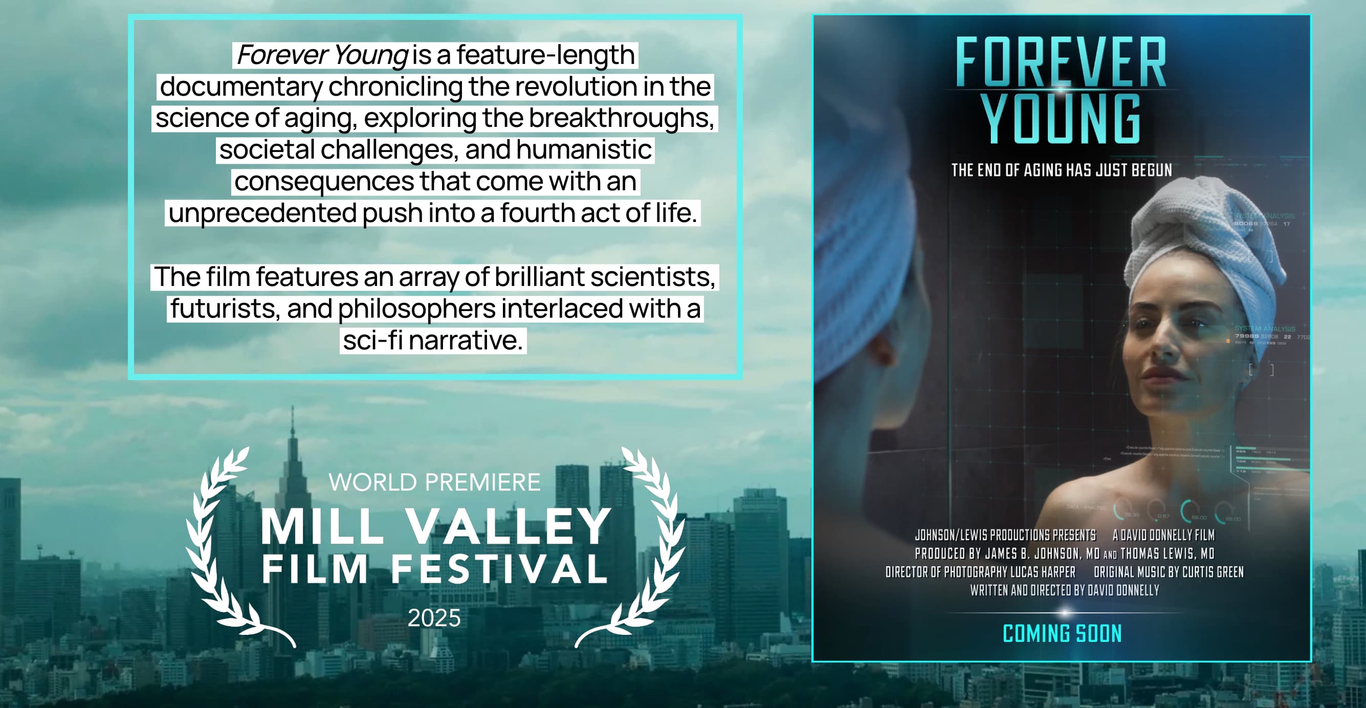
So, I was very happy to recently see this new movie. I think this film is the ideal resource. It translates complex biological developments into accessible and compelling content, making it perfect for sharing with those outside the field.

So I encourage you to do what I’m doing… Invite a few friends over, or sit down with your family and kids, and introduce them to the new world of healthy longevity! Or rent the movie and share the link and password with your friends or family members to get them up to speed on this exciting field.

The cost is only $12.95 US for a 7-day rental, without geographical limitations.

Click on this “Forever Young” link to watch the movie => Forever Young

Full disclosure: I will get some portion of the proceeds from the movie. But, it is by far the best movie I’ve seen on this topic, and I’m using it as a method to recruit more people into a longevity lifestyle.

If you want to learn more about the background of the movie, there was a good story this week in “Longevity Technology” here: Forever Young explores the longevity revolution

RapAdmin

This article examines the recent surge in comprehensive reports from global institutions—including the World Economic Forum, the OECD, and major financial entities—focusing on the profound economic implications of increasing human life expectancy. The central thesis is that longevity is no longer merely a public health success story but a dominant macroeconomic force that necessitates a fundamental restructuring of societal systems.

The summary highlights three primary themes:

  1. The Workforce Paradigm Shift: As populations age and birth rates decline, the traditional three-stage life model (education, work, retirement) is becoming obsolete. Reports suggest that “multi-stage lives” will require individuals to work longer, necessitating continuous lifelong learning and more flexible career paths. Companies are urged to eliminate ageism and adapt to intergenerational workforces to maintain productivity.
  2. Financial Sustainability and the “Longevity Economy”: The article notes a shift in how financial institutions view aging. Rather than seeing the elderly solely as a “silver tsunami” of rising costs, there is an increasing focus on the “longevity economy”—the economic contribution of people over 50. However, this requires urgent reforms in pension systems and wealth management to prevent widespread poverty in extended old age.
  3. Policy and Infrastructure Gaps: There is a significant disconnect between the reality of longer lives and current policy frameworks. The article calls for “longevity-ready” cities, healthcare systems that prioritize prevention over acute care, and social policies that support caregivers.

The author concludes that while the “longevity revolution” offers immense potential for human flourishing, the window for proactive economic adjustment is closing. Success depends on whether leaders treat aging as a looming crisis to be managed or a structural transformation to be embraced through innovation in education, finance, and labor.

Read the full article: It’s Raining Longevity Reports — As Age Hits Economies (Forbes)

Recent Reports:

RapAdmin

Sarcopenia—the age-associated loss of skeletal muscle mass, quality, and function—is a primary driver of frailty, metabolic dysregulation, and all-cause mortality in older adults. Standard clinical interventions rely on resistance training and high-protein nutrition, which frequently yield diminishing returns in advanced age due to cellular anabolic resistance and mitochondrial decay. Recent data identifies a targeted botanical intervention capable of bypassing this resistance to rescue muscle decline at the molecular level.

Researchers investigated the therapeutic potential of Peucedanum japonicum (PJ), a medicinal botanical, and its primary bioactive metabolite, 4-caffeoylquinic acid (4-CQA). To test efficacy in a clinically relevant model of aging, the team administered the compound to 20-month-old mice—an age roughly biologically equivalent to 60-year-old humans—for an eight-week period. The results demonstrated a robust reversal of sarcopenic progression.

The core mechanism of action is the restoration of mitochondrial respiratory capacity. Aging skeletal muscle is characterized by the accumulation of damaged mitochondria and an involuntary shift from powerful fast-twitch (Type II) to endurance-based slow-twitch (Type I) muscle fibers. The administration of PJ and 4-CQA upregulated PGC-1alpha, the master transcriptional coactivator of mitochondrial biogenesis. This activation sequentially triggered the Nrf1-TFAM signaling axis, stimulating the transcription of nuclear and mitochondrial genes required to construct new, high-functioning electron transport chain complexes. Simultaneously, the intervention halted muscle catabolism. It effectively suppressed the ubiquitin-proteasome system by downregulating the expression of critical atrogenes (Atrogin-1 and MuRF1), which are responsible for dismantling contractile proteins during aging and disuse. Macroscopically, treated mice exhibited increased lean body mass, expanded muscle cross-sectional area, restored fast-twitch fiber ratios, and significantly enhanced functional output in both grip strength and forced treadmill endurance.

For longevity optimization, this positions 4-CQA beyond the scope of a generic antioxidant. It acts as a targeted metabolic exercise mimetic, chemically stimulating the mitochondrial pathways typically reserved for rigorous physical training, thereby offering a practical pharmacological strategy to preserve mobility and extend healthspan.

Source:

RapAdmin

A critical translation of preclinical longevity data into human neurodegeneration has materialized in a six-month, single-arm pilot trial evaluating weekly rapamycin administration in patients with early-stage Alzheimer’s disease (AD). Targeting the mechanistic target of rapamycin (mTOR) pathway, which regulates cellular growth and autophagy, this intervention aims to alter the natural trajectory of cognitive and metabolic decline.

The primary finding is an unexpected stabilization of brain glucose metabolism. In typical AD progression, patients exhibit distinct metabolic declines in specific cortical regions. However, following 26 weeks of an intermittent 7 mg oral rapamycin dose, participants demonstrated no significant reduction in [18F]FDG uptake within the prespecified primary regions: the posterior cingulate cortex, precuneus, and temporoparietal lobe. Furthermore, exploratory regional analyses identified significant metabolic increases in the putamen, insula, and anterior cingulate cortex, contradicting the established hypometabolic trajectory of the disease.

Simultaneously, the trial generated highly paradoxical cerebrospinal fluid (CSF) biomarker data. Treatment induced significant elevations in total tau, neurofilament light chain (NfL), and amyloid-beta 40, alongside a numerical increase in amyloid-beta 42. While typically interpreted as an exacerbation of neuroaxonal damage, phosphorylated tau (p-tau) remained largely stable. This resulted in a significantly decreased p-tau/total tau ratio, suggesting the protein efflux may reflect enhanced autophagy-mediated clearance (secretory autophagy) rather than acute neurotoxicity.

The safety profile of weekly rapamycin in this geriatric, cognitively impaired demographic was acceptable. No serious adverse events were recorded; common toxicities were restricted to expected mild gastrointestinal disturbances and aphthous ulcers. Despite the lack of a placebo control, the data establish essential feasibility, target engagement, and safety groundwork for adequately powered, randomized controlled trials to evaluate mTOR inhibition as a disease-modifying strategy in neurodegeneration.

Source:
Pre-print paper: Evaluation of rapamycin as a neuroprotective treatment in Alzheimer’s disease: a six-month single-arm open-label clinical pilot trial
Institution: Karolinska Institutet and Karolinska University Hospital. Country: Stockholm, Sweden. Journal Name: medRxiv.

Leitura Relacionada: Rapamycin as a preventive intervention for Alzheimer’s disease in APOE4 carriers: targeting brain metabolic and vascular restoration

RapAdmin

The biological narrative of aging has traditionally viewed mitochondria as cellular powerhouses that slowly degrade over time, producing reactive oxygen species that passively damage the cell. However, a compelling synthesis of recent research positions mitochondria as active, albeit inadvertent, instigators of systemic “inflammaging.” The core mechanism centers on mitochondrial DNA (mtDNA) escaping its highly regulated matrix and acting as a potent signaling molecule—specifically, a damage-associated molecular pattern (DAMP).

Under physiological stress, mitochondrial quality control mechanisms fail. This leads to the release of mtDNA into the cytosol or extracellular space via mitochondrial-derived vesicles (MDVs), membrane pores (BAX/BAK, VDAC, mPTP), or complete cellular rupture. Because mtDNA is of alphaproteobacterial origin and lacks typical nuclear methylation patterns, the innate immune system recognizes it as a foreign, pathogen-like invader.

Once free, mtDNA triggers a cascade of inflammatory pattern recognition receptors (PRRs). In the endosomes, it activates Toll-like receptor 9 (TLR9); in the cytosol, it is detected by the cGAS-STING pathway, and inflammasomes like NLRP3, AIM2, and ZBP1. This recognition initiates the production of type I interferons and pro-inflammatory cytokines (IL-1beta, IL-18), driving sterile inflammation that accelerates cardiovascular aging, atherosclerosis, and heart failure. Furthermore, circulating cell-free mtDNA serves as a vector for inter-organ cross-talk, transmitting inflammatory distress signals from a failing heart to the kidneys, lungs, and brain.

The identification of mtDNA as a primary driver of chronic aging pathologies provides highly actionable targets. Interventions must focus on three pillars: stabilizing mitochondrial membranes to block mtDNA release, inhibiting downstream PRR sensors, or, most practically for longevity biohackers, upregulating selective autophagy (mitophagy) to clear damaged mitochondria before they leak.

Source:

Leitura Relacionada:

RapAdmin

Researchers from the University of Minnesota and the University of Georgia have identified a new class of senolytics that exploit an inherent vulnerability in senescent cells: their susceptibility to ferroptosis. Traditionally, senolytic drugs—such as the dasatinib and quercetin combination—have been designed to induce apoptosis by disrupting senescent cell anti-apoptotic pathways (SCAPs). However, these compounds often function as broad-spectrum anticancer agents with significant off-target toxicities. This study shifts the paradigm by demonstrating that conjugated polyunsaturated fatty acids (PUFAs), specifically α-eleostearic acid (α-ESA) and its methyl ester derivative (α-ESA-me), selectively clear senescent cells through an entirely different mechanism.

Senescent cells accumulate high levels of labile ferrous iron and reactive oxygen species (ROS). They also upregulate key enzymes in the arachidonic acid metabolism pathway, including Acyl-CoA synthetase long-chain family member 4 (ACSL4), lysophosphatidylcholine acyltransferase 3 (LPCAT3), and arachidonate 15-lipoxygenase (ALOX15). When introduced to this primed environment, conjugated PUFAs like α-ESA act as highly oxidizable fuel. The ACSL4-LPCAT3-ALOX15 enzymatic axis incorporates these lipids into cellular membranes, where they undergo rapid, iron-dependent lipid peroxidation. This massive accumulation of lipid hydroperoxides overwhelms the cell’s glutathione peroxidase 4 (GPX4) detoxification capacity, leading to catastrophic membrane damage and ferroptotic cell death.

In vivo testing revealed that short-term administration of α-ESA-me in naturally aged wild-type mice reduced senescence markers across multiple tissues, including the liver, kidney, and lungs. Furthermore, chronic administration in an Ercc1-/Δ progeroid mouse model improved composite healthspan scores (e.g., tremor and kyphosis reduction) and decreased tissue senescence.

Source:

RapAdmin

Geroscience has historically focused on extending lifespan and “healthspan”—the years free from debilitating chronic disease. However, a new perspective paper argues this framework is deeply flawed because it overlooks the gradual erosion of capacity that occurs even in ostensibly healthy individuals. The researchers introduce “Peakspan”: the period during which an individual maintains at least 90% of their peak functional performance in a specific physiological or cognitive domain. According to their multi-system analysis, most human biological systems reach maximal capacity in early adulthood, between the 20s and 30s.

Consequently, humans now spend the vast majority of their adult lives in a “healthy but declined” state, carrying a massive functional gap well before clinical disease is diagnosed.

The authors argue that extending Peakspan, rather than just healthspan, is the true functional manifestation of rejuvenative biomedical progress and is strictly necessary for sustained economic growth in aging societies. They advocate for a massive shift in how we measure and intervene in aging, leveraging artificial intelligence (AI) and “Deep Aging Clocks”. Instead of measuring chronological age deviation, these AI models would calculate a “delta-peak age,” estimating an individual’s proximity to their own historical maximum performance rather than a population average. By aggressively targeting the specific moment when function drops below 90% (the “Peakspan exit”), we can theoretically intervene before system-wide decline cascades into overt pathology. The paper essentially demands we stop accepting gradual functional deterioration as “normal” and start utilizing AI to optimize for maximal capacity throughout the life course.

Source: