Notícias

RapAdmin

Gut Check: Common Postbiotic ‘Butyrate’ Identified as Potent Senomorphic

Researchers at the University of Birmingham report that butyrate, a short-chain fatty acid made by fibre-fermenting gut bacteria, can blunt the ageing of human T cells. In older people, both stool and blood butyrate levels fall, and lower levels track with more “senescent” (worn-out but inflammatory) T cells. In the lab, adding butyrate to activated T cells from older donors dialled down their inflammatory secretions (IL-6, IL-8, IL-1beta), reduced DNA-damage and mitochondrial stress markers, and suppressed mTOR/NF-kB signalling. In mice, transferring butyrate-rich stool fluid from young animals cut the build-up of senescent T cells in the spleen. The work positions butyrate as a candidate “senomorphic” — a drug that quiets senescent cells rather than killing them.

As we age, a population of exhausted immune cells accumulates in the body. These “senescent” T cells stop dividing but refuse to die quietly — instead they leak a cocktail of inflammatory molecules that drives the chronic, smouldering inflammation (“inflammaging”) linked to everything from Alzheimer’s to heart failure and rheumatoid arthritis. Clearing or silencing these cells is one of the hottest strategies in longevity medicine.

A team led by Niharika Duggal at the University of Birmingham, working with the Quadram Institute, has now pointed the finger at the gut. The big idea: the trillions of microbes in our intestines help keep the immune system young by producing short-chain fatty acids — chiefly butyrate — when they ferment dietary fibre. As the microbiome ages and diversity is lost, butyrate production falls, and the immune system may pay the price.

The researchers first showed the correlation in people. Comparing 40 young (18-37) and 40 older (60+) adults, they found butyrate dropped with age in both stool and, for the first time, in blood serum. Crucially, the less butyrate an older person had in their stool, the more senescent T cells circulated in their blood.

To test causation, they turned to the lab. Taking T cells from older donors and forcing them to divide repeatedly — an accelerated ageing model — they bathed some in butyrate. The treated cells behaved younger: they secreted far less IL-6 and IL-8, carried fewer DNA-damage marks, generated less mitochondrial “rust” (reactive oxygen species), and showed damped activity in the mTOR and NF-kB pathways that orchestrate inflammatory ageing. A gene-expression scan confirmed butyrate switched down a signature panel of senescence and inflammation genes.

Finally, in aged mice whose microbiomes had been wiped with antibiotics, a transplant of butyrate-rich fluid from young mouse stool reduced the number of senescent T cells and inflammatory IL-6 in the spleen.

The authors are careful: this is mechanistic and preclinical work, not a clinical trial. But it strengthens the case that a high-fibre diet, prebiotics, or eventually butyrate supplements could become a gentle way to keep the ageing immune system in check. The next step is a proper human trial.

Actionable Insights

The practical hook is dietary, not pharmaceutical — the paper itself recommends raising butyrate “through dietary, microbial, and therapeutic approaches,” specifically more fibre (fruit, vegetables, legumes, whole grains), prebiotics (inulin, fructooligosaccharides), and butyrate-producing probiotics. Supplement forms floated for future trials are oral sodium butyrate and the precursor tributyrin. No dose, formulation, or human efficacy for anti-ageing has been established, so any action today is a reasonable bet on general gut health, not a proven longevity intervention.

Effect-size reality check: the strongest human data point is a correlation — stool butyrate explained about 43% of the variance in senescent CD8 T-cell frequency (R-squared = 0.43, equivalent to r ≈ 0.66, a large association; and R-squared = 0.21, r ≈ 0.46 for a second subset). This is an association, not proof that raising butyrate lowers senescence in you.

RapAdmin

A team of researchers at Cornell University has identified a specific molecular “coating” on the surface of embryonic stem cell-derived (EVs) that appears to be the master switch for their potent anti-aging effects. While the scientific community has long known that “young” EVs can rejuvenate “old” cells, the precise mechanism has remained a “black box”—until now.

In a study published in October 2025, lead author Shun Enomoto and senior author Richard Cerione demonstrated that EVs shed by mouse embryonic stem cells (mESCs) are not merely sacks of genetic cargo, but are coated externally with the extracellular matrix protein fibronectin. This fibronectin coating is the critical “key” that unlocks the antioxidant defenses of recipient cells. Upon contact with aging fibroblasts (skin cells) and astrocytes (brain cells), the fibronectin-coated vesicles bind to integrin receptors, triggering a kinase cascade (FAK/AKT) that stabilizes the “longevity factor” Nrf2. This effectively rewires the aging cell to neutralize oxidative stress and escape senescence.

Impact Evaluation: The Journal of Biological Chemistry has an Impact Factor of approximately 3.9–4.0 (2024/2025 data). Evaluated against a typical high-end range of 0–60+ for top general science, this is a High impact journal (historically the “workhorse” of rigorous mechanistic biochemistry), though not an “Elite” broad-interest outlet like Nature or Cell. Its reputation for technical rigor validates the specific signaling pathway identified.


The Biohacker Analysis

Study Design Specifications

  • Type: In Vitro (Cell Culture mechanistic study).
  • Subjects:
    • Source: Mouse Embryonic Stem Cells (mESCs).
    • Targets: Mouse Embryonic Fibroblasts (MEFs) and Astrocytes.
    • Controls: EVs from differentiated cells (non-stem) or Fibronectin-depleted EVs.

Organ Priority: Skin (Fibroblasts) and Brain (Astrocytes).

Novelty

The “Dogma Shift” here is the focus on the corona (surface protein coating) rather than the lumen (internal cargo). Most exosome startups are obsessed with what is inside the vesicle (miRNAs). This paper suggests that the mere contact of a fibronectin-coated vesicle with a cell is sufficient to trigger the Nrf2 longevity response, even before the cargo is unloaded.

Critical Limitations

  • No In Vivo Data: The study does not show that injecting these EVs into old mice extends life. It only shows that it keeps cells “young” in a dish. This is a massive translational gap.
  • Tumorigenicity Risk: Embryonic stem cells (ESCs) are associated with teratoma formation. While EVs are cell-free, the growth-promoting signals (AKT activation) they carry are pro-proliferative. Activating AKT and Nrf2 in a pre-cancerous cell could theoretically accelerate tumor growth.
  • Species Gap: Verified in mouse cells. Human validation is the required next step.

Actionable Intelligence

The Translational Protocol

Warning: This study uses a complex biologic (EVs), not a simple drug. The following extrapolations apply to “Exosome Therapy” and “Nrf2 Activators.”

  • Human Equivalent Dose (HED) for Exosome Therapy:

    • Based on standard mouse-to-human EV scaling (typically 100 µg protein/mouse):
    • Math: 100 µg / 0.02 kg mouse = 5 mg/kg mouse dose.
    • HED: $5 \text{ mg/kg} \times (3/37) \approx 0.4 \text{ mg/kg}$.
    • For a 70kg Human: ~28 mg of purified Exosome protein (intravenous).
    • Note: Commercial “exosome” clinics often under-dose significantly compared to this academic standard.
  • Pharmacokinetics (PK/PD):

    • Bioavailability: EVs injected IV have a half-life of 2–30 minutes in circulation before being cleared by the liver/spleen/lungs. However, their signaling effects (like Nrf2 activation) can last for hours to days.
    • Targeting: Fibronectin-coated EVs may naturally home to injured tissues expressing specific integrins.
  • Safety & Toxicity Check:

    • NOAEL: Not established for Embryonic EVs in humans.
    • Red Flag: AKT Activation. Chronic activation of AKT is a known driver in many cancers. “Pulsing” this therapy is safer than chronic elevation.
    • Immunogenicity: Allogenic (donor-derived) EVs can trigger immune responses, though less than whole cells.

Biomarker Verification Panel

  • Efficacy Markers:
    • Nuclear Nrf2: Hard to measure clinically.
    • Downstream Targets: Increase in GSH (Glutathione) levels and HO-1 (Heme Oxygenase-1) protein in PBMCs (blood cells).
    • Senescence Load: Reduction in p16INK4a expression in T-cells.
  • Safety Monitoring:
    • Cancer Screen: Because this pathway (AKT/Nrf2) protects cells from dying, it can also protect cancer cells. Strict annual cancer screening (Liquid Biopsy/Galleri test) is mandatory if using potent Nrf2 activators or growth factors.

Feasibility & ROI

  • Sourcing: Not Available. You cannot buy “Fibronectin-coated Embryonic EVs” legally for human use.
  • The “Hack”: The study identifies Nrf2 as the goal.
    • Alternative: Sulforaphane (Broccoli sprout extract) is a validated dietary Nrf2 stabilizer, though it works via Keap1 inhibition, not the FAK/AKT axis.
    • Alternative: Astaxanthin helps preserve mitochondrial function and may crosstalk with this pathway.
  • Cost: High-end “Exosome Therapy” in offshore clinics costs $5,000–$15,000 per session. The ROI is Low until human efficacy is proven; the risk-to-reward ratio is poor compared to standard small molecules.

Population Applicability

  • Contraindications: Active Cancer. Do not activate Nrf2 or AKT if you have a tumor; it protects the tumor from oxidative stress (chemo/radiation resistance).

The Strategic FAQ

  1. Q: If I can’t get Embryonic EVs, can I just take Fibronectin supplements?

    • A: No. Fibronectin is a massive protein that would be digested in the gut. Even if injected, it needs to be presented on a membrane surface (like an EV) to cluster integrins correctly to fire the FAK signaling signal. Free-floating fibronectin does not have the same effect.
  2. Q: Does this confirm that “Young Blood” (Parabiosis) works via exosomes?

    • A: It strengthens the case. This paper aligns with previous work (e.g., Nature Aging 2024) showing young plasma EVs extend life. It adds the specific mechanism (Fibronectin/Nrf2) that was previously vague.
  3. Q: Is Nrf2 activation always good?

    • A: No. “Nrf2 addiction” is a phenomenon in cancer cells where they use Nrf2 to survive chemotherapy. You want cyclic activation (pulsed) to clear senescent cells or repair damage, not chronic activation which might promote malignancy.
  4. Q: How does this compare to Rapamycin (mTOR inhibition)?

    • A: They are opposite but complementary. Rapamycin slows growth and induces autophagy (cleaning mode) by inhibiting mTOR. These EVs activate AKT (growth mode) and Nrf2 (defense mode). You wouldn’t want to do both simultaneously; they might cancel each other out. A cycling protocol (Growth vs. Repair) would be the theoretical approach.
  5. Q: Are commercial “Exosome” IVs in clinics the same as this?

    • A: No. Commercial exosomes usually come from Mesenchymal Stem Cells (MSCs) from umbilical cords or fat. This study used Embryonic Stem Cells (ESCs). The protein coating and cargo are likely different. We do not know if MSC-exosomes use this specific Fibronectin-Nrf2 pathway.
  6. Q: What is the “half-life” of the anti-aging effect?

    • A: In the dish, the effect “potently blocked” senescence during the stress window. In vivo, because EVs are cleared in minutes, the signaling ripple effect likely lasts 24–48 hours. This suggests a need for frequent dosing, which makes it financially unfeasible for most.
  7. Q: Can I boost my own endogenous stem cell EVs?

    • A: Likely yes, via exercise and fasting, which mobilize stem cells. However, as you age, your stem cells (and their EVs) degrade. The paper implies you need embryonic (pristine) EVs to get the full “reset.”
  8. Q: Did they check for teratomas (tumors) in this study?

    • A: Data Absent. The study was in vitro (cell culture). Teratoma risk is a primary concern with any embryonic-derived product and must be ruled out in future mouse studies.
  9. Q: Is there a small molecule that mimics this FAK-AKT-Nrf2 axis?

    • A: There are FAK inhibitors (for cancer), but few specific FAK activators. This highlights why biologics (EVs) can do things small molecules can’t—they can structurally cluster receptors to turn on a complex pathway.
  10. Q: What is the “Kill Switch” if this therapy goes wrong?

    • A: There isn’t one. Once you inject exosomes, they distribute systemically. This is why safety trials for ESC-derived products are incredibly slow and rigorous.

Source Paper (Open Access): Embryonic stem cell-derived extracellular vesicles delay cellular senescence by inhibiting oxidative stress

Leitura Relacionada: Forever young? Extracellular vesicles may be key to halt aging | Cornell Chronicle

RapAdmin

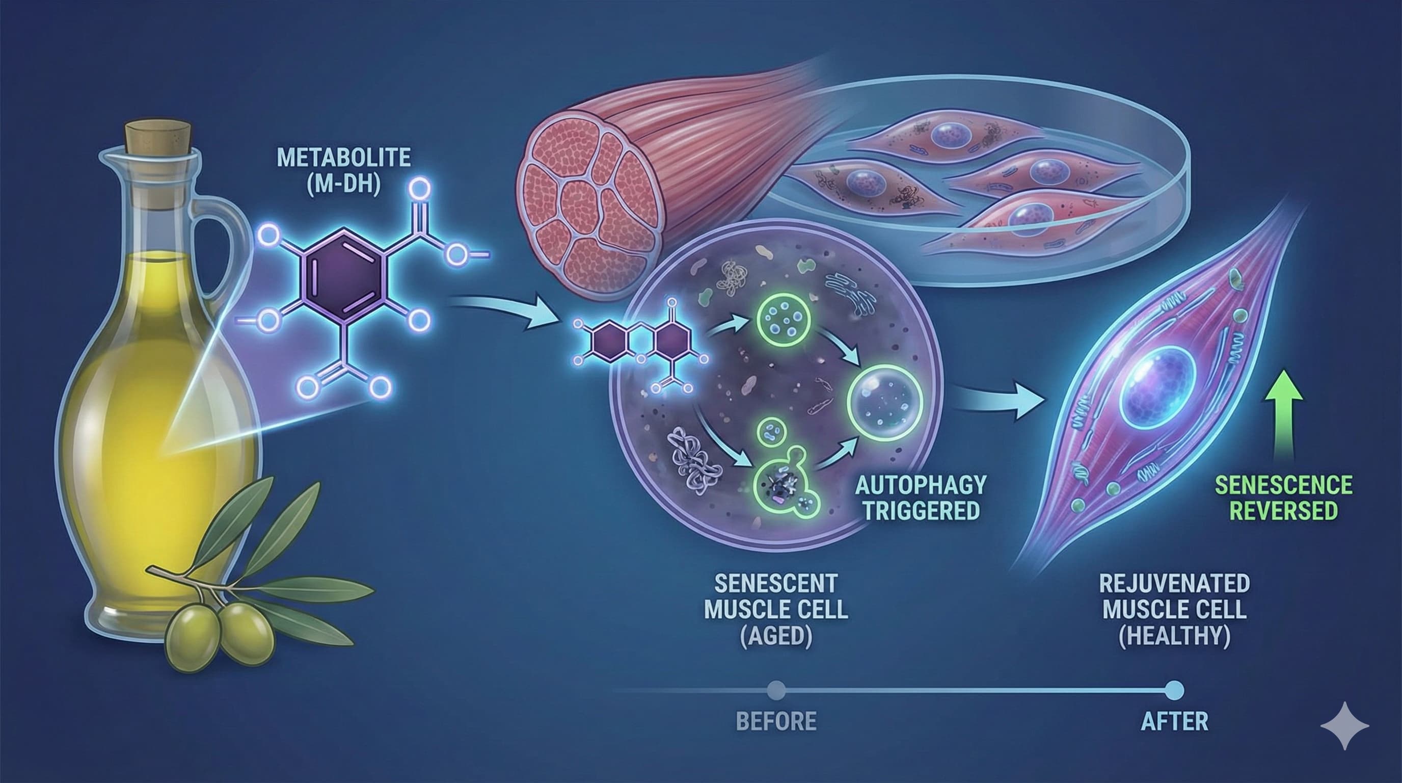
In a significant addition to the “Food as Medicine” pharmacopeia, researchers from the University of Udine (Italy) and Sorbonne Université (France) have elucidated the specific molecular mechanics by which Oleuropein Aglycone (OLE)—a bioactive metabolite found in Extra Virgin Olive Oil (EVOO)—protects human skeletal muscle from oxidative aging. Published in BioFactors, this study moves beyond generic “antioxidant” claims, identifying OLE as a potent activator of the AMPK-FOXO3a-Sestrin axis.

The “Big Idea” here is the identification of a nutritional intervention that directly targets sarcopenia (age-related muscle loss) at the cellular level. By treating human immortalized myoblasts (AB1079) with OLE prior to oxidative assault (H2O2), the team observed a 43% reduction in reactive oxygen species (ROS) and a 12% reduction in cellular senescence. Crucially, OLE didn’t just scavenge free radicals; it turned on the cell’s internal cleaning crew (autophagy) and mitochondrial quality control systems. While this is an in vitro study, it provides the mechanistic validation needed to prioritize high-phenolic olive extracts in longevity protocols, specifically for maintenance of lean muscle mass in the elderly.

Context:

  • Institution: University of Udine (Italy), Sorbonne Université (France).
  • Journal: BioFactors (Wiley).
  • Impact Evaluation: The impact score of this journal is ~5.4 (JIF 2024), evaluated against a typical high-end range of 0–60+ for top general science, therefore this is a High impact journal (Q1/Q2 in Biochemistry/Endocrinology).

Study Design Specifications

  • Type: In vitro (Cellular Biology).
  • Subjects: Human skeletal muscle cells (Immortalized myoblast cell line AB1079).
  • Lifespan Data: N/A. This study did not perform lifespan analysis on organisms (e.g., mice or C. elegans). It measured cellular survival and senescence markers.
  • Dose/Concentration: Cells were treated with Oleuropein Aglycone (OLE) prior to H2O2 exposure. (Specific micromolar concentrations were used to determine IC50 and efficacy).

Mechanistic Deep Dive

The paper identifies Oleuropein Aglycone (OLE) not merely as a direct antioxidant, but as a xenohormetic signal that activates longevity pathways:

  1. AMPK Activation: OLE induces phosphorylation of AMPK, the cell’s “energy sensor,” mimicking the effects of fasting or exercise.
  2. FOXO3a Translocation: Activated AMPK triggers FOXO3a to enter the nucleus, upregulating stress-resistance genes.
  3. Sestrin Upregulation (SESN2/SESN3): This is the novel finding. OLE specifically upregulates Sestrins, which act as a scaffold to further activate AMPK and suppress mTORC1, preventing cellular senescence.
  4. Autophagy Restoration: The treatment restored autophagic flux (clearing damaged organelles) which is typically blunted by oxidative stress.
  • Organ-Specific Priority: Skeletal Muscle. The mechanism suggests utility in preventing sarcopenia and maintaining metabolic flexibility.

Novelty

We knew olive oil was “good” for you. We didn’t know exactly how it preserved muscle tissue under stress. This paper links the polyphenol OLE directly to the Sestrin-AMPK feedback loop in human muscle cells, providing a specific genetic target (SESN2/3) that explains the preservation of muscle function observed in Mediterranean populations.

Critical Limitations

  • No In Vivo Translation: This is a cell-culture study. There is no guarantee that oral ingestion of OLE achieves the tissue concentrations used in the petri dish.
  • Metabolic Stability: The study uses Oleuropein Aglycone, which is a metabolite. Most supplements contain the glycoside (Oleuropein). The conversion rate in vivo is variable and microbiome-dependent.
  • Single Cell Line: Results in AB1079 myoblasts may not perfectly mirror primary muscle fibers in an aged human body with systemic inflammation.

RapAdmin

This research paper is from 2021, but I thought it would be of interest to people here:

Broccoli’s Master Molecule Reverses Skin Aging at the Genetic Level

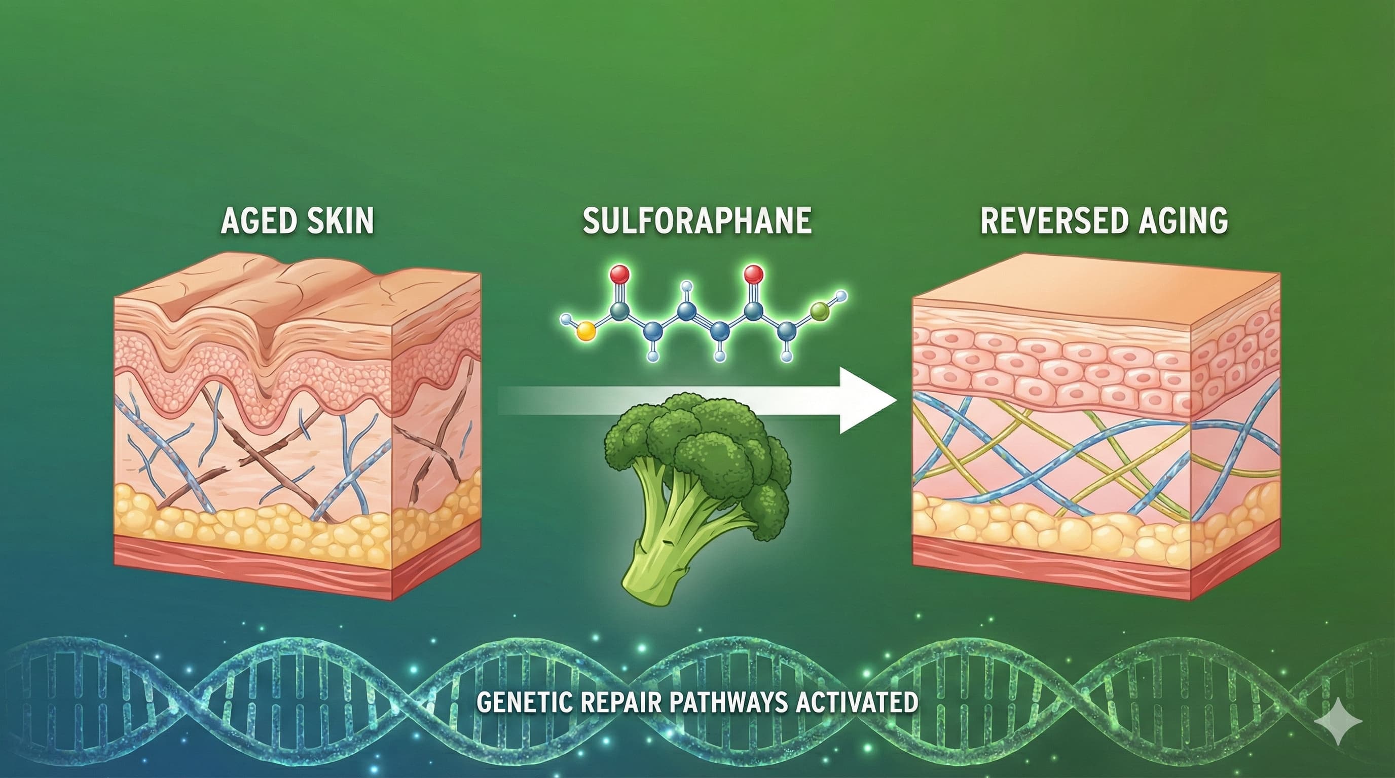
In a compelling demonstration of “food as medicine,” researchers have reversed biological markers of skin aging in mice using a high-dose dietary regimen of Sulforaphane (SFN), the potent bioactive compound found in broccoli. While the cosmetic industry obsesses over topical creams, this study reveals that systemic, internal activation of the Keap1-Nrf2 pathway—the body’s master antioxidant switch—can physically restructure the dermal layer from the inside out.

The study compared young (2-month) and old (21-month) mice, finding that the elderly cohort suffered from collapsed collagen networks and high oxidative stress. However, when these “geriatric” mice were fed a diet fortified with SFN for three months, their skin’s genetic profile began to mimic that of youth. The treatment successfully restored collagen deposition and upregulated critical defense genes (like NQO1 and HO1) that usually go dormant with age. Crucially, this wasn’t just about reducing inflammation; it was a structural remodeling of the extracellular matrix, suggesting that longevity compounds could make our largest organ—the skin—functionally younger, not just smoother.

Context:

  • Institution: University of Coimbra (Portugal) & Texas Tech University Health Sciences Center (USA).
  • Journal: Journal of Nutritional Biochemistry.
  • Impact Evaluation: The impact score of this journal is ~4.9 (2024 JIF), evaluated against a typical high-end range of 0–60+ for top general science, therefore this is a Medium impact journal. (While not Nature or Science, it is a respected, specialized tier-1 journal in nutritional science).

Part 2: The Biohacker Analysis

Study Design Specifications

  • Type: In vivo (Animal Study).
  • Subjects: Male C57BL/6 mice.
    • Groups: Young (2 months) vs. Old (21 months).
    • Treatment: Sulforaphane (SFN) incorporated into the diet at 442.5 mg/kg of diet for 3 months.
  • Lifespan Data: Not Measured. The study was terminated after 3 months to harvest tissue. No longevity survival curves were generated.

Mechanistic Deep Dive

The study targets the Nrf2 (Nuclear factor erythroid 2-related factor 2) pathway, often called the “guardian of longevity.”

  • The Problem: In aging skin, Nrf2 gets trapped by a protein called Keap1 and degraded, preventing it from entering the nucleus to activate defense genes. This leads to ROS (Reactive Oxygen Species) accumulation and collagen degradation via MMP9.
  • The Fix: SFN modifies Keap1 cysteine residues, liberating Nrf2.
  • Key Findings:
    • Gene Expression: Old treated mice showed a massive resurgence in antioxidant mRNA (Nrf2, NQO1, HO1), restoring levels comparable to young mice.
    • Collagen: Masson’s trichrome staining revealed that SFN significantly improved collagen structural integrity in old mice.
    • Mitochondrial/ROS: Protein levels of ROS and MMP9 (an enzyme that chews up collagen) were significantly suppressed.

Novelty

Most skin aging studies focus on topical application (creams/serums). This paper validates systemic, dietary intervention for dermal remodeling. It proves that skin aging is a metabolic issue that can be addressed centrally via the gut-skin axis, specifically utilizing the Nrf2 pathway to reverse established structural damage, not just prevent it.

Critical Limitations

  • Supra-Physiological Dosing: The dose used (442.5 mg/kg diet) translates to a massive human equivalent dose (see Part 3). This is likely unachievable via eating broccoli alone.
  • No Functional Lifespan Data: We do not know if these mice lived longer, only that their skin looked better.
  • Dermal Thickness Gap: While collagen quality improved, the actual thickness of the dermis in old mice did not fully recover to young levels, suggesting a “ceiling” to the regenerative effect.
  • Gender Bias: Only male mice were used. Estrogen plays a massive role in skin aging (collagen production); excluding females is a major translational blind spot.

Part 3: Actionable Intelligence

The Translational Protocol (Rigorous Extrapolation)

WARNING: The dose extrapolated below is considered HIGH and potentially toxic for humans if taken as a pure isolate. Proceed with extreme caution.

1. Human Equivalent Dose (HED) To translate the mouse dose to humans, we use Body Surface Area (BSA) normalization factors (Reference: FDA Guidance for Industry).

  • Mouse Input: 442.5 mg SFN per kg of diet.
  • Daily Intake Estimate: A standard mouse consumes approximately 150g of food per kg of body weight (BW) per day.
    • Math: 442.5 (mg/kg diet) x 0.15 (kg food/kg BW) = 66.4 mg/kg BW/day (Mouse Dose).
  • HED Calculation:
    • Formula: Mouse Dose / 12.3 (Conversion Factor).
    • Math: 66.4 / 12.3 = 5.4 mg/kg BW.
  • For a 75kg Human:
    • Math: 5.4 x 75 = ~405 mg/day.
  • Context: Most clinical trials for autism or prostate cancer use 15–60 mg/day. A 405 mg dose is roughly 6x to 10x higher than standard therapeutic doses.

2. Pharmacokinetics (PK/PD)

  • Bioavailability: Sulforaphane has high absolute bioavailability (~80%) but a very short half-life.
  • Half-Life: Approximately 1.8 to 2.5 hours in humans. (Source: PubMed - Pharmacokinetics of SFN).
  • Implication: To mimic the continuous dietary intake of the mice, a human would need Pulsed Dosing (e.g., 100mg every 6 hours). Single daily dosing is insufficient for sustained Nrf2 activation.

3. Safety & Toxicity Check

  • Toxicity Signals: In rat toxicity studies, sedation, hypothermia, and motor impairment appear at doses >150 mg/kg. The calculated HED (5.4 mg/kg) is below the lethal threshold but enters the range of potential hepatotoxicity and gastrointestinal distress. (Source: ToxNet / PubChem Data).
  • Interactions: SFN is a potent inhibitor of CYP3A4. It may alter the metabolism of statins, benzodiazepines, and immunosuppressants.

4. Biomarker Verification Panel

  • Efficacy: Do not rely on “feeling better.” Test GGT (Gamma-Glutamyl Transferase)—low levels correlate with good glutathione status. Advanced users can test Urinary Mercapturic Acids (SFN-NAC) to verify absorption.
  • Safety Monitoring: Liver Panel (ALT/AST) is mandatory at these doses. Watch for thyroid suppression (goitrogenic effect of high-dose crucifers).

5. Feasibility & ROI

  • Sourcing: Pure “Stabilized Sulforaphane” is rare and expensive. Most supplements are “Glucoraphanin + Myrosinase” (precursor blends).
  • Cost Estimate:
    • To hit ~400mg SFN using high-end supplements (e.g., Broc Shot @ 12mg/dose) would require ~33 doses/day.
    • Cost: >$2,000/month.
    • Verdict: Economically Unfeasible for this specific study protocol. Users must rely on lower, optimized doses (e.g., 30-50mg) combined with synergistic Nrf2 activators (Curcumin, Resveratrol) to achieve similar effects.

Source Research Paper (Open Access): Dietary supplementation with sulforaphane ameliorates skin aging through activation of the Keap1-Nrf2 pathway

RapAdmin

If there is a “silver bullet” in the longevity pharmacopeia, this review confirms it is Rapamycin. In a field often plagued by hype and unreplicable results, the inhibition of the Mechanistic Target of Rapamycin (mTOR) stands apart as the only intervention with a near-perfect batting average across every model organism tested. This 2025 review serves as a definitive dossier, arguing that we have moved past asking if mTOR inhibition works, and must now focus on how to deploy it for maximum benefit in humans.

The authors detail how hyperactive mTOR signaling is the central engine of aging—driving cellular senescence (“zombie cells”) and blocking repair. By inhibiting this pathway, Rapamycin flips a genetic switch from “growth at all costs” to “maintenance and repair,” effectively mimicking the profound benefits of calorie restriction without the metabolic misery. Most critically for humans, the data shows that intervention in late life is still effective, reversing age-related decline even when started in the biological equivalent of a 60-year-old. The message is clear: It is not too late to slow the clock.

Source Review Paper:

  • Institution: Shoolini University, VSB-Technical University of Ostrava, et al. (International Consortium).
  • Country: India / Czech Republic / Slovakia.
  • Journal Name: EXCLI Journal.
  • Impact Factor: 4.9 (Significant impact in experimental clinical sciences).

Part 2: The Biohacker Analysis

Study Design Specifications

  • Type: Systematic Review & Meta-Analysis of the “Gold Standard” literature.
  • Subjects: Complete evolutionary spectrum: Yeast (S. cerevisiae), Worms (C. elegans), Flies (D. melanogaster), and Mice (M. musculus).
  • Lifespan Data (The “Upside”):
    • Yeast: Chronological lifespan extended by ~54%.
    • Worms: Median lifespan extension of ~19%.
    • Flies: Females show up to 23% maximum lifespan extension.
    • Mice: The most compelling data for humans.
      • Consistency: Reproducible extension of 10-18% across genetically diverse strains (unlike other compounds that fail in heterogeneous groups).
      • Late-Onset Efficacy: Treatment started at 20 months (old age) still extended remaining lifespan significantly.

Mechanistic Deep Dive: The Engine of Youth

The review confirms that mTOR inhibition is not merely slowing damage accumulation; it is actively restoring youthful cellular function through three key pathways:

  1. Restoration of Autophagy:
  • The Mechanism: mTORC1 naturally inhibits autophagy. Rapamycin releases this brake.
  • The Benefit: This triggers the cellular “clean-up crew,” digesting misfolded proteins (Alzheimer’s plaques) and dysfunctional mitochondria. This is the primary driver of the anti-aging effect.

  1. Senomorphic Activity:
  • The Mechanism: Instead of just killing senescent cells (senolytics), mTOR inhibition suppresses their ability to secrete inflammatory signals (the SASP).
  • The Benefit: This dramatically reduces systemic inflammation (“inflammaging”) without destroying the tissue architecture.
  1. Stem Cell Preservation:
  • The Mechanism: By preventing stem cell exhaustion (hyper-proliferation), rapamycin maintains the regenerative pool.
  • The Benefit: Enhanced wound healing and tissue maintenance in the long term, contrary to the fear that it stops growth.

Novelty

This paper solidifies the transition of Rapamycin from an “immunosuppressant” to an “immunomodulator.” The authors present data showing that while high doses suppress immunity, longevity-associated doses actually rejuvenate the immune system in the elderly (likely by clearing senescent immune cells), improving response to vaccines and infection—a massive paradigm shift.

Critical Limitations (The Engineering Challenge)

  • The Dosage Puzzle: The “toxicity” often cited is a result of continuous dosing (transplant protocols). The paper implies, but does not solve, that the key to avoiding side effects is pulsed dosing.
  • Glucose dysregulation: The review notes hyperglycemia in some models, but newer interpretations suggest this may be “starvation diabetes” (a benign physiological shift to fat oxidation) rather than pathological insulin resistance.

Part 3: Actionable Intelligence

The Protocol: The “Bull Case” Translation

  • Strategy: High-Dose Intermittent (Pulse) Therapy.
  • Rationale: Maximize mTORC1 inhibition (autophagy trigger) while allowing mTORC2 recovery (safety) during the off-days.
    • Protocol: 5mg – 8mg of Rapamycin (Sirolimus) taken once weekly.
    • Absorption Hack: Take with a high-fat meal (e.g., olive oil, avocado) to increase bioavailability by up to 30%, getting more “bang for your buck.”
    • The “Stack”: Pair with Acarbose (to blunt glucose spikes) or Glycine (to support methylation).

Biomarkers: Optimization Metrics

Don’t just look for toxicity; look for efficacy:

  1. Inflammation: Target an hs-CRP < 0.5 mg/L. Rapamycin is a potent anti-inflammatory; a drop here is a sign of success.
  2. Biological Age Clocks: If available (DNAmGrimAge), track the deceleration of epigenetic aging.
  3. HOMA-IR: Monitor insulin resistance. If it creeps up, adjust the pulse frequency (e.g., once every 10 days instead of 7) rather than quitting.

Feasibility & ROI

  • Cost-Benefit: Extreme Value. Generic Sirolimus is relatively inexpensive ($1-$3 per mg).
  • ROI: Compared to unproven supplements (NMN, Resveratrol) which cost hundreds monthly for marginal/placebo gains, Rapamycin offers the highest probability of genuine biological age reversal for the dollar.

Population Applicability

  • Broad Spectrum: Unlike interventions that require specific genetic makeups, the conservation of the mTOR pathway implies this will work for the vast majority of humans.
  • Target Demographic: Ideally suited for individuals 40+, where natural autophagy begins to decline precipitously.

Source Paper (Open Access): Exploring the role of mTOR pathway in aging and age-related disorders

RapAdmin

New research from York University in Toronto challenges the prevailing view of how our muscles age, identifying a specific “garbage disposal” failure that exercise can reverse. For years, longevity science focused on mitophagy—the tagging of damaged mitochondria for removal—as the primary bottleneck in muscle aging. However, a comprehensive review published in Experimental Physiology by Anastasiya Kuznyetsova and Dr. David A. Hood suggests the problem isn’t identifying the trash, but destroying it.

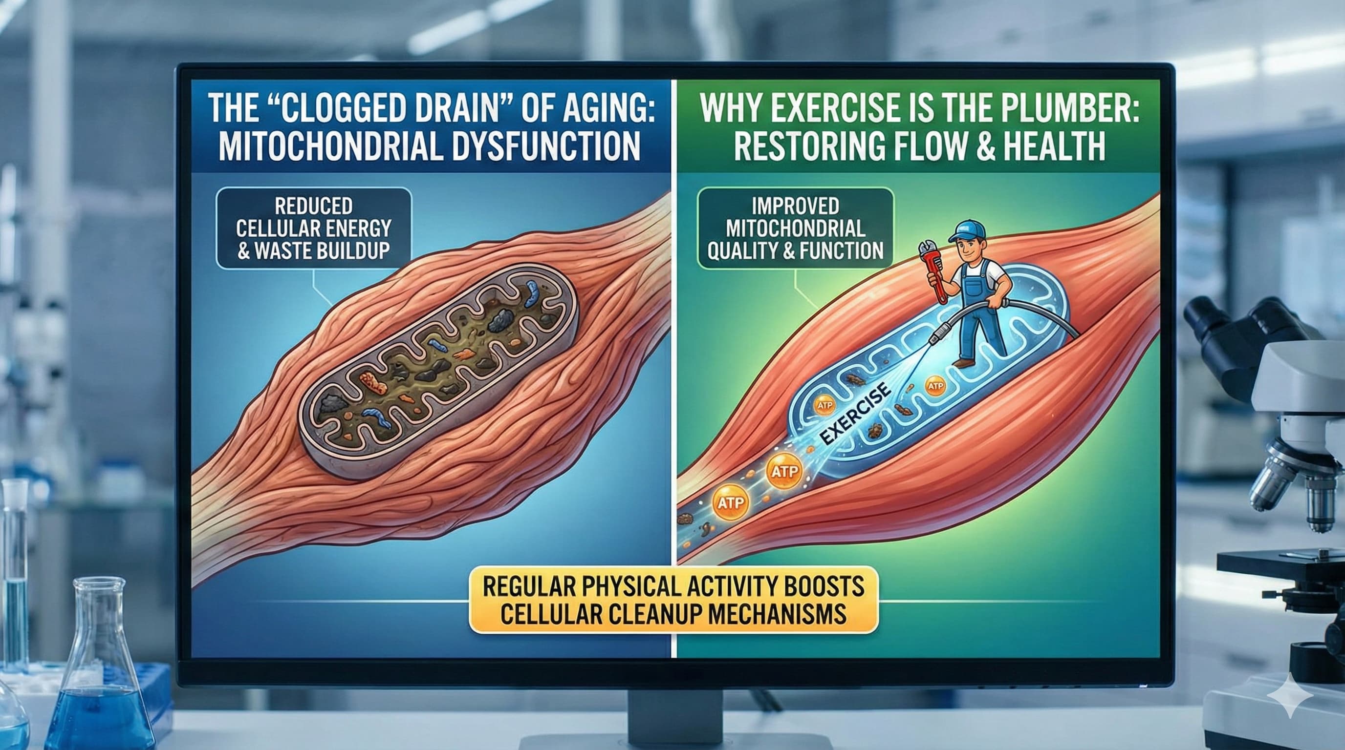
The study highlights that aging skeletal muscle is not starved of mitophagy signals; in fact, it is often overflowing with them. Aged muscle cells show a paradoxical increase in lysosomal proteins (the cellular incinerators) and mitophagy markers. The critical failure occurs at the terminal step: the lysosomes themselves become “clogged” with undigested cellular debris (lipofuscin) and lose their proteolytic (protein-chopping) acidity. This effectively turns the muscle cell into a hoarding situation where marked trash accumulates because the incinerator is broken.

Crucially, the researchers detail how specific forms of exercise—specifically chronic endurance-style contractile activity—act as a lysosomal “reboot.” Exercise doesn’t just stimulate the creation of new mitochondria (biogenesis); it restores the acidity and enzymatic efficiency of lysosomes, allowing them to finally clear the backlog of dysfunctional organelles. This finding shifts the target for longevity interventions from simply “triggering autophagy” to “restoring lysosomal acidity,” offering a more precise roadmap for preventing sarcopenia and maintaining metabolic flexibility into late life.


Mechanistic Interpretation

  • The Bottleneck is Downstream: The paper argues that upstream signaling (AMPK activating ULK1, or PINK1/Parkin tagging mitochondria) often remains intact or even hyperactive in aging as a compensatory mechanism.
  • Lysosomal dysfunction: The core pathology is a loss of lysosomal proteolytic capacity (likely due to rising pH or enzyme deactivation) and the accumulation of lipofuscin (indigestible protein aggregates), which physically blocks further degradation.
  • TFEB Regulation: Exercise activates Transcription Factor EB (TFEB), the master driver of lysosome biogenesis, which not only builds more lysosomes but “refreshes” the enzymatic pool, restoring the ability to degrade cargo.

Novelty

  • Paradigm Shift: Contradicts the idea that aging is purely a “deficiency” of autophagy signals. Instead, it frames aging as a “traffic jam” or “indigestion” where the cell tags waste faster than it can degrade it.
  • Hyper-accumulation: The finding that lysosomal proteins (like LAMP1) and mitophagy markers increase with age (2-fold in some models) represents a failed compensatory response, not a successful adaptation.

Actionable Insights for Biohackers

  • Prioritize Lysosomal pH: Interventions should focus on acidifying lysosomes, not just “boosting autophagy.”
    • Stack: Consider Spermidine (a possible lysosomal activator) alongside exercise.
    • Mimetics: Urolithin A is highly relevant here for its specific ability to stimulate mitophagy, but it must be paired with lysosomal support to prevent “trash accumulation.”
  • Exercise Dosing:
    • Modality: The study emphasizes chronic endurance/aerobic work (Zone 2) as the primary driver of lysosomal remodeling. Resistance training drives hypertrophy but is less effective at this specific “cleaning” mechanism.
    • Volume: Consistency is key. The “clogged drain” clears slowly; sporadic high-intensity bouts may not be sufficient to restore enzymatic pools.
  • Biomarkers to Watch:
    • Lipofuscin: Hard to measure directly in vivo without biopsy, but skin accumulation (age spots) can be a proxy for systemic lysosomal health.

Cost-Effectiveness

  • ROI: High. Zone 2 cardio is free. The mechanistic benefit (clearing cellular junk) prevents downstream metabolic collapse, which is far more expensive to treat.
  • Supplementation: Low-to-Moderate. Basic lysosomal support (fasting/Zone 2) is free. Advanced mimetics (Urolithin A) are expensive ($100+/month) and should only be added if the “free” foundation is solid.

Critical Limitations

  • Model Reliance: Much of the mechanistic “clogging” data is derived from rodent models (e.g., specific mouse strains). Human lysosomal kinetics are slower and harder to measure.
  • Translation Gap: The “dose” of exercise required to reverse lysosomal pH drift in an 80-year-old human is currently unknown.
  • Invasiveness: The gold-standard metrics (lysosomal pH, lipofuscin load) require muscle biopsies, making n=1 tracking difficult for biohackers.

Publication Details:

RapAdmin

The binary view of cellular aging—that a cell is either healthy or “senescent”—is increasingly viewed as a dangerous oversimplification. New research from the University of Tasmania and Monash University, Australia, published in the top-tier journal Aging Cell, suggests that “zombie cells” exist on a pathogenic spectrum, and that the longevity drug rapamycin works by specifically targeting the most toxic sub-populations among them.

Using high-throughput single-cell fluorescence imaging on human fibroblasts, the researchers demonstrated significant heterogeneity in classical senescence biomarkers, including p21, SA-βgal, and IL-6. They discovered that nuclear morphology is a robust predictor of toxicity; cells with enlarged nuclei were highly correlated with elevated Interleukin-6 (IL-6) expression, a primary driver of the Senescence-Associated Secretory Phenotype (SASP). This confirms that not all senescent cells contribute equally to “inflammaging.”

Mechanistically, the study reveals that rapamycin does not act as a blanket senolytic (killing all senescent cells). Instead, it functions as a precision senomorphic. The drug selectively dampened the high-intensity biomarkers in the specific sub-populations characterized by enlarged nuclei and hyper-active SASP. This implies that rapamycin’s longevity benefits may stem from modulating the mTOR pathway to suppress the protein synthesis required for the toxic secretory phase of specific “super-senescent” cells, rather than simply halting the cell cycle or inducing autophagy universally. By silencing the “loudest” cells responsible for systemic inflammation, rapamycin effectively lowers the cumulative inflammatory burden without requiring total senescent cell clearance.

Actionable Insights for Longevity Biohackers

  • Refining the Rapamycin Protocol: The data supports the “senomorphic” use case for rapamycin. Rather than high-dose “shock” therapy intended to kill cells, consistent, modulation-focused dosing (e.g., weekly pulsed dosing) may be more effective at keeping the high-SASP sub-populations suppressed, preventing the systemic inflammatory cascade.
  • Morphological vs. Chemical Biomarkers: Standard commercial senescence tests often rely on aggregate SA-βgal. This study suggests this is insufficient. Biohackers conducting n=1 trials should prioritize circulating inflammatory markers (hs-CRP, IL-6, TNF-α) as proxies for the activity of senescent cells, rather than just their presence.
  • Vascular Health Proxy: Given that fibroblasts (connective tissue) were the model, monitoring pulse wave velocity (arterial stiffness) or endothelial function could serve as a functional readout for whether the suppression of fibrotic/senescent signaling is translating to tissue flexibility.

Cost-Effectiveness

  • ROI: High. Rapamycin (sirolimus) is a generic pharmaceutical. Given that this study highlights its ability to neutralize the most damaging subset of senescent cells without the need for expensive, novel senolytics, the marginal benefit per dollar is exceptional compared to proprietary peptides or experimental gene therapies.

Critical Limitations

  • In Vitro Constraints: The study utilized cultured human fibroblasts induced via chemotherapy (mitomycin C) and oxidative stress. This ignores the complex immune surveillance (NK cells, macrophages) present in a live organism that usually clears these cells.
  • Senomorphic vs. Senolytic Ambiguity: While the study shows biomarker suppression, it does not confirm if these “tamed” cells eventually undergo apoptosis or if they rebound immediately upon cessation of rapamycin, necessitating lifelong adherence.
  • Tissue Specificity: Fibroblast behavior may not translate to post-mitotic tissues like neurons or cardiomyocytes. The nuclear-enlargement correlation might be specific to structural cells.

Source Research Paper (Open Access): Single-Cell Fluorescence Imaging Reveals Heterogeneity in Senescence Biomarkers and Identifies Rapamycin-Responsive Sub-Populations (Aging Cell) Sept. 25

RapAdmin

The modern pursuit of longevity is defined by a central tension: the trade-off between anabolic growth (necessary for muscle mass and performance) and catabolic repair (necessary for clearing cellular debris and extending lifespan). This dichotomy is governed by the Mechanistic Target of Rapamycin (mTOR). The prevailing dogma has long suggested an “Interference Effect”: that inhibiting mTOR for longevity via rapamycin would catastrophically blunt the adaptive response to exercise, rendering the biohacker frail and metabolically stagnant.

This report provides a critical analysis of the landmark study “Rapamycin does not prevent increases in myofibrillar or mitochondrial protein synthesis following endurance exercise” by Philp et al. (2015). This research fundamentally challenges the interference dogma. It offers mechanistic proof that the mammalian system can uncouple the signaling pathways for muscle hypertrophy from those for mitochondrial biogenesis.

For the longevity biohacker, this distinction is critical. It implies that while rapamycin may dampen muscle size gains (hypertrophy) for a period after dosing, it does not impair—and may potentially augment—the metabolic improvements associated with endurance training. This analysis breaks down the study’s methodology, explores the “PGC-1α compensatory surge,” and outlines a translation-focused protocol for integrating rapamycin with endurance training without sacrificing metabolic fitness.

The Discovery of Rapamycin and the mTOR Pathway

To understand the significance of the Philp et al. study, one must view it through the lens of evolutionary biology. mTOR is an evolutionarily conserved serine/threonine kinase that acts as the master nutrient sensor.

  • Nutrient Abundance: When amino acids (leucine) and growth factors (IGF-1) are present, mTOR Complex 1 (mTORC1) activates, driving ribosome biogenesis and protein synthesis.
  • Nutrient Scarcity: When nutrients are low, mTORC1 is inhibited. This triggers a survival program: the cell halts growth and initiates autophagy, a catabolic process where the cell recycles damaged organelles. This induction of autophagy is the primary mechanism behind the lifespan-extending effects of rapamycin.

The Interference Hypothesis

For the active biohacker, mTOR presents a paradox. Exercise is a potent activator of mTORC1 in skeletal muscle, driving recovery and adaptation. The logical inference, known as the “Interference Hypothesis,” was that taking rapamycin (an mTOR inhibitor) would block the benefits of exercise.

If rapamycin prevents the cell from sensing “growth” signals, does it also prevent the cell from building new mitochondria or repairing muscle fibers? The Philp et al. (2015) study serves as the definitive test of this hypothesis in the context of endurance exercise, dissecting whether “growth” and “metabolic adaptation” are inextricably linked.

Biohacking Synthesis – Actionable Insights

Based on the Philp et al. data, we can construct a “Rapamycin-Endurance” protocol that leverages the uncoupling effect.

The “Uncoupling” Protocol

The goal is to inhibit mTOR for longevity (autophagy) without sacrificing metabolic fitness (VO2 max, mitochondrial density).

  • Modality Selection: Prioritize Zone 2 / Steady State Endurance. The study confirms that mitochondrial adaptations are preserved under rapamycin. Avoid high-intensity hypertrophy work (heavy lifting) during the peak therapeutic window of rapamycin, as MyoPS is blunted.
  • Dosing Timing:
    • The “Fast-Mimetic” Stack: Take rapamycin 1-2 hours before a long, low-intensity endurance session. The Philp data suggests this will not block mitochondrial biogenesis and may augment PGC-1α signaling.
    • The “Growth” Separation: To preserve muscle mass, perform resistance training as far as possible from the rapamycin dose (e.g., dosing on a rest day or 24-48 hours post-dose), as human mTOR inhibition may last longer than in mice.
  • Biomarker Tracking:
    • Functional: Monitor VO2 Max and Zone 2 watts/kg. If these decline, the mitochondrial uncoupling may not be fully translating to your specific physiology.
    • Structural: Monitor Appendicular Lean Mass (ALM) via DEXA. If muscle mass drops, the blunting of MyoPS is too severe, and protein intake or timing must be adjusted.

Cost-Effectiveness and ROI

Rapamycin rates highly for cost-effectiveness in the longevity pharmacopoeia.

  • Generic Availability: Sirolimus is widely available as a generic.
  • Cost: Off-label prescriptions typically cost $1.00–$3.00 per mg. A standard weekly protocol (e.g., 6mg) costs roughly $25–$35 per month.
  • Relative ROI: Compared to peptide therapies or proprietary supplements, rapamycin offers one of the highest “mechanism-per-dollar” ratios due to the robustness of the lifespan data and the low monthly cost.

Source Research Paper (Open access): Rapamycin does not prevent increases in myofibrillar or mitochondrial protein synthesis following endurance exercise

Gemini In-depth analysis of paper: https://gemini.google.com/share/6d57280d8393

Leitura Relacionada: Rapamycin & Muscle Growth Decoupled: Longitudinal Hypertrophy Bypasses mTOR

RapAdmin

The Silent Burn: How Systemic Inflammation Dismantles the Brain’s Hydraulic Infrastructure

In a paradigm-shifting review published in Experimental Gerontology , researchers from China Medical University have re-framed the etiology of “Cognitive Frailty”—the dangerous intersection of physical weakness and cognitive decline. Moving beyond the neuron-centric models that have dominated Alzheimer’s research for decades, the authors propose a “vascular-inflammatory axis” as the primary driver of this condition. The central thesis is stark: the aging brain does not simply wither; it starves, choked off by a collapsing microvascular network that has been degraded by chronic, systemic inflammation.

The paper, titled “Chronic inflammation and cognitive frailty in older adults: A narrative review” (Jiang et al., 2025), synthesizes emerging evidence that pro-inflammatory cytokines—specifically IL-6, TNF-α, and CRP—are not merely passive markers of aging but active agents of structural destruction. These molecules, originating from senescent cells and visceral fat, launch a relentless assault on the body’s microcirculation, the delicate web of capillaries responsible for nourishing tissues. In the brain, this results in the breakdown of the Blood-Brain Barrier (BBB), neurovascular uncoupling, and eventually, the simultaneous manifestation of sarcopenia (muscle loss) and cognitive impairment.

For the longevity community and biohackers, this report serves as a critical biological signal: preserving cognitive function is isomorphic with preserving endothelial integrity. The authors detail mechanistic pathways, including the cGAS-STING axis—a cellular alarm system triggered by leaking mitochondrial DNA—that drives this sterile inflammation. Crucially, the review posits that this state is reversible. Interventions ranging from the Dietary Inflammatory Index (DII) optimization to specific mind-body exercises like Baduanjin are highlighted as potent levers to dampen this inflammatory fire and restore microvascular perfusion. The implication is profound: to save the neuron, one must first save the vessel.

The Sources of Sterile Inflammation

Jiang et al. identify several upstream sources of the cytokines driving CF. These sources are critical targets for biohacker interventions because they represent the “root causes” of the downstream damage.

  • Cellular Senescence (The SASP): As cells accumulate DNA damage or reach their replicative limit (Hayflick limit), they enter a zombie-like state called senescence. They do not die, but they stop dividing. Crucially, they become metabolically hyperactive, secreting a toxic cocktail of pro-inflammatory cytokines (IL-6, IL-1β, IL-8), chemokines, and proteases. This is the Senescence-Associated Secretory Phenotype (SASP). In CF, endothelial cells lining the blood vessels become senescent, turning the vasculature itself into a source of inflammation.
  • Visceral Adipose Tissue: Fat is not merely energy storage; it is an endocrine organ. In aging and metabolic syndrome, visceral fat becomes infiltrated with macrophages (M1 phenotype) that secrete high levels of TNF-α and IL-6. This “adipose inflammation” spills over into the systemic circulation, reaching the brain and muscle tissues.
  • Gut Dysbiosis (“Leaky Gut”): The review and supporting literature point to the gut-brain axis. With age, the intestinal barrier permeability increases, allowing bacterial endotoxins (Lipopolysaccharides, LPS) to enter the bloodstream. This condition, metabolic endotoxemia, triggers a systemic immune response, chronically elevating CRP levels and driving neuroinflammation.
  • Immunosenescence: The aging immune system loses its ability to distinguish “self” from “non-self” and to clear pathogens effectively. This results in a dual failure: a weakened response to new infections (like flu) but a heightened, non-specific autoimmune-like activity against the body’s own tissues.

The Neural Consequences

When these cytokines cross the Blood-Brain Barrier (or damage it sufficiently to enter), they activate the brain’s resident immune cells: the microglia. In a healthy brain, microglia are housekeepers, pruning synapses and clearing debris. In an inflamed brain, they adopt a “primed” or aggressive phenotype. They stop cleaning and start attacking, releasing reactive oxygen species (ROS) and excitotoxins (like glutamate) that kill neurons. This “neuroinflammation” is the biological bridge linking systemic inflammation to the cognitive deficits seen in CF.

Continue reading the full story in the next post, here: Interventions and Protocols: Actionable Insights

Source Paper (Open Access): Chronic inflammation and cognitive frailty in older adults: A narrative review

Gemini Analysis of the paper: https://gemini.google.com/share/11560ad80357