Notícias

RapAdmin

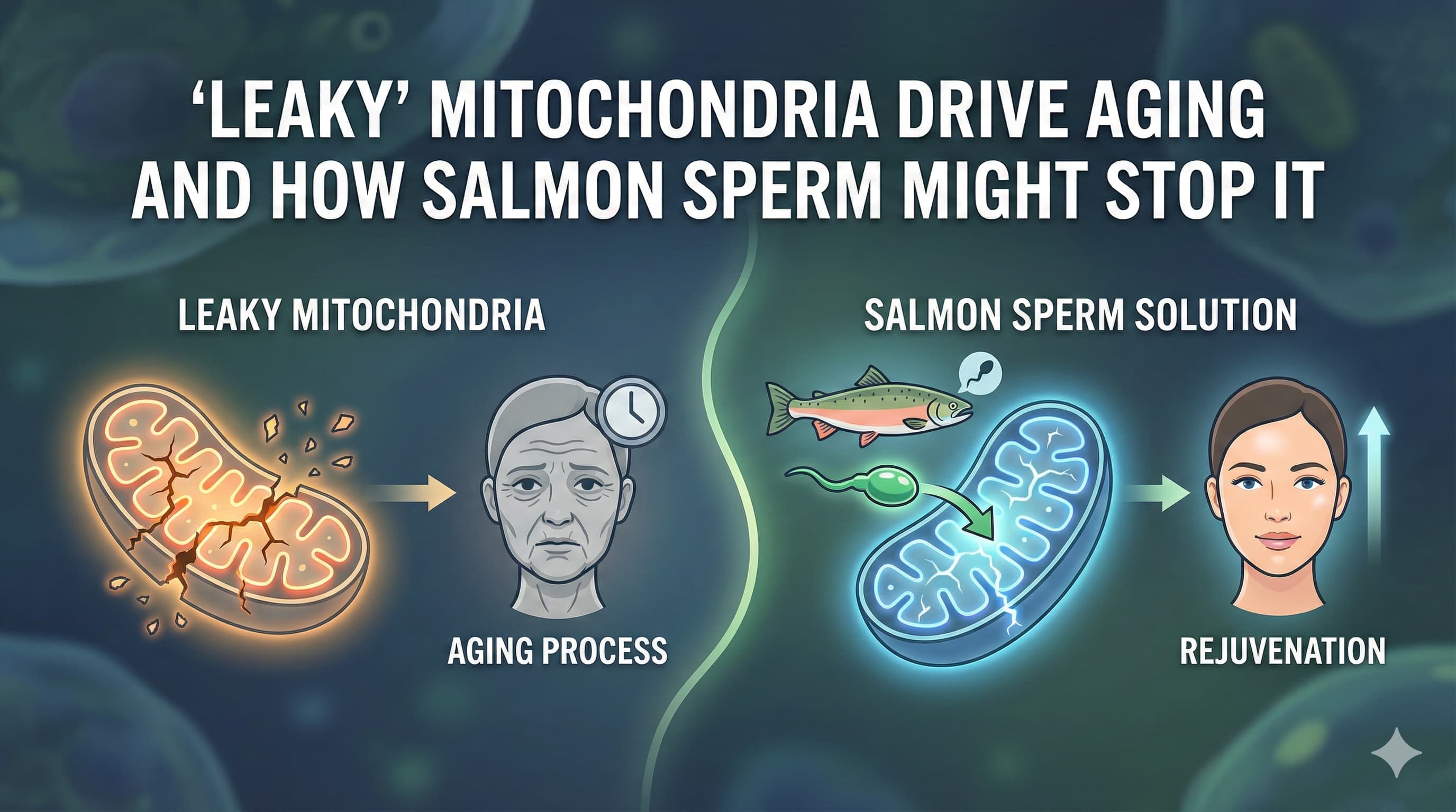
A paradigm-shifting study published in Nature (September 2025) by researchers at the Max Planck Institute for Biology of Ageing (Germany) has identified a “hidden” cause of age-related inflammation: the accidental incorporation of RNA building blocks into mitochondrial DNA (mtDNA). This molecular error, caused by a metabolic supply chain failure in aging cells, renders the mitochondrial genome structurally unstable. The fragile DNA subsequently leaks into the cell’s cytosol, triggering a false alarm in the innate immune system (cGAS-STING) that drives the chronic, low-grade inflammation known as “inflammaging.”

The research team, led by Amir Bahat and Dusanka Milenkovic under the supervision of Thomas Langer, discovered that senescent cells stop producing sufficient deoxyribonucleotides (dNTPs)—the specific “bricks” required for DNA—because they arrest their cell cycle and downregulate the enzyme ribonucleotide reductase (RNR). However, their mitochondria continue to replicate. Starved of the correct “deoxy” building blocks, the mitochondrial polymerase is forced to utilize abundant RNA building blocks (rNTPs). This “contaminated” mtDNA is chemically fragile and prone to breakage.

Crucially, the researchers demonstrated that this process is reversible. By supplementing cells with deoxyribonucleosides (dNs)—the specific precursors to DNA found in high concentrations in sources like salmon milt—they restored the metabolic balance. This intervention prevented mtDNA fragility, halted the leakage, and suppressed the inflammatory Senescence-Associated Secretory Phenotype (SASP). This establishes a direct mechanistic link between nucleotide metabolism and the immune drivers of aging, suggesting that “refueling” the mitochondrial DNA synthesis machinery with exogenous DNA precursors could be a potent, cost-effective longevity intervention.

Translational Biohacking: The Deoxyribonucleoside Intervention

The most actionable finding of the Bahat et al. paper for the n=1 experimenter is that exogenous supplementation with deoxyribonucleosides (dNs) suppresses the phenotype. By feeding the cells the specific precursors they lack, the researchers restored the dNTP pool, excluded ribonucleotides from mtDNA, prevented leakage, and silenced the inflammatory signal.

4.1 The “Nucleotide” Confusion: RNA vs. DNA Supplements

This is the most critical distinction for any biohacker attempting to replicate this protocol. The dietary supplement market is flooded with “Nucleotides,” but these are almost exclusively RNA-based.

  • Standard Nucleotides: Derived from Yeast (Saccharomyces cerevisiae). These are high in RNA. Hydrolysis yields ribonucleosides (Adenosine, Guanosine, Cytidine, Uridine). Supplementing with these would increase the rNTP pool, potentially worsening the rNTP:dNTP ratio imbalance that causes the problem.
  • Required Nucleotides: Derived from DNA. The study explicitly calls for deoxyribonucleosides (Deoxyadenosine, Deoxyguanosine, Deoxycytidine, Thymidine).
  • Thymidine vs. Uridine: A key marker. RNA uses Uridine; DNA uses Thymidine. If a supplement label lists “Uridine Monophosphate,” it is an RNA source. If it lists “Thymidine,” it is a DNA source.

Why it matters: The senescent cell has plenty of ribonucleotides; it is drowning in them. To fix the ratio, one must supplement the denominator (dNTPs) without increasing the numerator (rNTPs). You must consume DNA, not RNA.

4.2 The Source: Salmon Milt (The DNA Superfood)

Where does one find concentrated DNA in nature? The answer is sperm (milt). Sperm cells are essentially DNA delivery vehicles with very little cytoplasm (and thus less RNA/protein relative to DNA).

  • Salmon Milt Extract (SME): In industrial biology and functional food markets, the primary source of deoxyribonucleotides is Salmon Milt (soft roe).
  • Composition: Commercial DNA extracts from salmon milt are approximately 90% DNA and very low in RNA.
  • Japanese Market: In Japan, “Salmon Milt DNA” (Shirako) is a recognized functional food and supplement, often marketed for liver health and anti-aging.
  • Comparison:
    • Yeast Extract: ~85% RNA, <5% DNA. (Incorrect tool).
    • Salmon Milt: ~90% DNA, <1% RNA. (Correct tool).

4.3 Pharmacokinetics: Can you eat DNA?

A common skeptical critique is that dietary DNA is digested into base sugars and not absorbed intact. While true that DNA is hydrolyzed, it is not biologically wasted.

  1. Digestion: Pancreatic nucleases (DNase I) break DNA down into nucleotides and nucleosides in the gut.
  2. Absorption: Phosphatases convert nucleotides to nucleosides (base + sugar), which are absorbed by intestinal transporters (CNT and ENT family transporters).
  3. The Salvage Pathway: Once in the blood, these deoxyribonucleosides are taken up by cells and phosphorylated back into dNTPs via the “Salvage Pathway” (using enzymes like Thymidine Kinase 2 - TK2 and Deoxycytidine Kinase - dCK).
  4. Bypassing RNR: Crucially, the salvage pathway bypasses the Ribonucleotide Reductase (RNR) enzyme. Even if the senescent cell has turned off RNR (the de novo pathway), it can still use the salvage pathway to build dNTPs if the raw nucleosides are provided in the blood.

Evidence of Efficacy:

  • Mouse Models: In mice with TK2 deficiency (a model of dNTP depletion similar to the senescent state), oral supplementation with deoxycytidine and thymidine extended lifespan from 13 days to 34+ days.
  • Human Safety: Clinical trials of Salmon Milt DNA (up to 12 weeks) showed safety and improvements in liver markers, proving biological activity in humans.10

See more here: Part 2, Actionable Insights for n=1 Experimentation

Source Research Paper (Open access): Ribonucleotide incorporation into mitochondrial DNA drives inflammation" by Bahat et al. (2025) in Nature

Gemini In-depth analysis of paper: https://gemini.google.com/share/6db4d16c6a0e

RapAdmin

In a paradigm-shifting study published in Nature Communications, researchers from the University of Chicago have exposed a treacherous flaw in the biological machinery of human skin—a mechanism where the body’s own cleaning systems are turned against it by sunlight. The research, titled “YTHDF2 regulates self non-coding RNA metabolism to control inflammation and tumorigenesis,” unravels a molecular tragedy that plays out in our cells every time they are exposed to ultraviolet B (UVB) radiation.

At the center of this drama is a protein called YTHDF2, a “reader” of the epigenetic code that normally acts as a guardian of cellular identity. Its job is to identify and destroy fragments of “self” RNA—specifically a molecule called U6 small nuclear RNA (snRNA)—that have been chemically modified with a marker known as N6-methyladenosine (m6A). Without YTHDF2, these RNA fragments accumulate and drift into the cell’s waste-processing centers, the endosomes. There, in a case of mistaken identity, the immune sensor Toll-like Receptor 3 (TLR3) confuses these harmless self-RNAs for viral invaders.

The University of Chicago team, led by Dr. Yu-Ying He and Dr. Chuan He, discovered that UVB radiation triggers a “kill switch” for YTHDF2. In a cruel twist, the radiation activates autophagy—the very process usually praised for extending life and cleaning cells—to specifically target and digest the YTHDF2 protein. With the guardian removed, the “viral mimic” U6 snRNA floods the system, triggering a massive inflammatory response that drives sunburn, tissue damage, and eventually, skin cancer.

This discovery rewrites the rulebook for longevity and dermatology. It suggests that the chronic inflammation of aging, or “inflammaging,” is not merely a passive accumulation of damage, but an active, runaway immune response triggered by a failure in RNA quality control. By identifying the specific molecular pathway—from the m6A tag to the lysosomal transporter SIDT2—the authors have illuminated new targets for intervention. If we can stabilize YTHDF2 or block the U6-TLR3 handshake, we might finally decouple the sun’s rays from the aging they cause, creating a biological “sunblock” that works from the inside out.

Actionable Insights for N=1 Experimentation

Disclaimer: These insights are theoretical and based on the reviewed literature. They do not constitute medical advice.

4.1 Biomarkers to Watch

To track the efficacy of interventions targeting the YTHDF2-TLR3 axis:

  • hs-CRP (High-sensitivity C-reactive Protein): The gold standard for systemic inflammation.
  • IL-6: A specific cytokine downstream of the TLR3-NF-κB pathway.
  • Erythema Response Time: A functional metric. Measure the time it takes for skin to redden under a constant UV source (e.g., natural noon sun). Increased resistance suggests maintained YTHDF2 integrity.
  • Ferritin: Often elevated in acute inflammation and macrophage activation.

4.2 The “Solar Shield” Protocol (Stacking)

Phase 1: Prevention (The “Armor”)

  • Topical Zinc Oxide: Non-negotiable. Blocks the UVB trigger.
  • Oral Nicotinamide (500mg BID): Proven to reduce new non-melanoma skin cancers and support DNA repair (PARP activity), acting synergistically with RNA protection.

Phase 2: Downstream Blockade (The “Dampener”)

  • Resveratrol (500mg) + Curcumin (500mg Phytosome): Both are potent inhibitors of NF-κB. They act as a firewall, preventing the signal from the endosome (TLR3) from reaching the nucleus.
  • Quercetin (400mg): Acts as a mild endosomal modulator and zinc ionophore, potentially altering the pH environment required for TLR3 signaling.

Phase 3: The Methylation Support (The “Tagging”)

  • TMG (Trimethylglycine) or Choline: Ensures ample supply of S-adenosylmethionine (SAMe), the methyl donor required by METTL3 to tag U6 snRNA with m6A. Without the tag, YTHDF2 cannot recognize and degrade the debris.

Phase 4: Autophagy Timing (The “Timer”) (Be Careful with Sun Exposure when dosing rapamycin)

  • Pulsed Rapamycin: If using rapamycin, take it in the evening or on days of low sun exposure. Taking a potent autophagy activator immediately before beach exposure is theoretically contraindicated by this study, as it could accelerate YTHDF2 loss.

4.3 Dosing Strategy

  • Circadian Alignment: The study implies YTHDF2 levels fluctuate. Support circadian biology (sleep, dark exposure) to maximize natural protein synthesis and repair cycles.
  • “Vacation Protocol”: During periods of intense UV exposure (vacations), discontinue autophagy activators (fasting, rapamycin) and switch to “building” (mTOR activation via protein) and “shielding” (antioxidants) to preserve protein mass, including YTHDF2.

Key Metrics:

  • Journal Rank: Nature Communications (Impact Factor ~16.6; Top-tier multidisciplinary journal).2
  • Authors’ Affiliation: University of Chicago, USA.2
  • Core Mechanism: UVB > YTHDF2 Dephosphorylation > Autophagic Decay > U6 snRNA Accumulation > Endosomal TLR3 Activation > Inflammation/Tumorigenesis.

Read Gemini’s full research paper analysis: https://gemini.google.com/share/b653054eb822

Source Paper (Open Access): YTHDF2 regulates self non-coding RNA metabolism to control inflammation and tumorigenesis (Nature Communications)

RapAdmin

The Pizza Herb that Mimics Fasting to Reverse Muscle Aging. A Poor-man’s Urolithin A

In a breakthrough study published in Nature Aging, researchers have identified Thymol—a common terpene found in thyme and oregano—as a potent activator of mitophagy (mitochondrial recycling) that significantly delays physiological aging.

The “Big Idea” here is Mitohormesis. The study reveals that Thymol acts as a mild mitochondrial toxin. It causes a “transient dampening” of the mitochondrial membrane potential. This slight bioenergetic stress signal (depolarization) tricks the cell into thinking its mitochondria are failing, which triggers the Pink1-Parkin pathway to clear out old, defective mitochondria (mitophagy) and build new ones.

The results are striking across multiple species: Thymol improved heat-stress survival in worms (C. elegans), prevented liver fat accumulation in obese mice, and—most importantly—slowed skeletal muscle decline and delayed epigenetic aging in senescence-accelerated mice. This suggests that your spice cabinet might hold a compound capable of mimicking the cellular benefits of exercise and fasting.

Impact Evaluation:
The impact score of Nature Aging is ~17.0 (JIF), evaluated against a typical high-end range of 0–60+ for top general science. Therefore, this is an Elite impact journal, ranking in the top tier of aging-focused publications alongside Cell Metabolism and Aging Cell.


Part 2: The Biohacker Analysis

Study Design Specifications

  • Type: In vivo (Zebrafish screen, C. elegans, Mice) and In vitro.
  • Subjects:
    • Zebrafish: High-content screening larvae.
    • C. elegans: Wild-type and pink-1 mutants (to verify mechanism).
    • Mice: Diet-Induced Obesity (DIO) C57BL/6J and Senescence-Accelerated Mouse Prone 8 (SAMP8).
  • Lifespan Data: The study focuses primarily on Healthspan and Epigenetic Aging rather than absolute median lifespan extension. Key findings include a significant “delay in epigenetic aging” (measured by DNA methylation clocks) and protection against sarcopenia (muscle loss).
  • Control Group: Vehicle (ethanol/DMSO) treated controls.

Mechanistic Deep Dive

  • Primary Target: Mitochondrial Membrane Potential (Delta-Psi-m).
  • Mechanism of Action: Thymol acts as a mild uncoupler. It induces a transient depolarization of the mitochondrial membrane.
  • Pathway: This drop in potential prevents Pink1 from being degraded. Stabilized Pink1 recruits Parkin, which tags damaged mitochondria for destruction (autophagy).
  • Validation: The effect was abolished in pink-1 deficient worms and cells, confirming the pathway is essential.
  • Organ Priorities:
    1. Skeletal Muscle: Reversal of sarcopenia/frailty.
    2. Liver: Prevention of steatosis (fatty liver) under high-caloric load.
    3. Epigenome: Reduction in biological age according to methylation clocks.

Novelty

We already knew polyphenols like Urolithin A induce mitophagy. The novelty here is the mechanism and the molecule.

  1. Thymol is a Terpene, not a Polyphenol: This opens a new chemical class for longevity.
  2. Mechanism is “Dirty” but Effective: Unlike “clean” receptor agonists, Thymol works by physically messing with mitochondrial bioenergetics (uncoupling). It proves that controlled toxicity (hormesis) is a viable longevity strategy.

Critical Limitations

  • The “Goldilocks” Problem: The mechanism relies on transient depolarization. Chronic, high-dose uncoupling is toxic (similar to the banned fat-burner DNP). The therapeutic window between “mitohormesis” and “energy failure” might be narrow.
  • Model Specificity: SAMP8 mice age artificially fast. Results in these mice are often more dramatic than in normal wild-type aging.
  • Bioavailability: Terpenes are volatile and often rapidly metabolized (glucuronidated) in the gut. The study likely used high doses to achieve tissue concentrations.

Part 3: Actionable Intelligence

The Translational Protocol (Rigorous Extrapolation)

Human Equivalent Dose (HED):

  • Animal Dose: Based on parallel studies (e.g., Kohlert et al.), effective mouse doses for thymol typically range from 20–50 mg/kg/day.
  • Calculation:
    • Formula: Mouse Dose (mg/kg) x (MouseKm / HumanKm)
    • Calculation: 40 mg/kg x (3 / 37) = 3.24 mg/kg.
  • Result: For a 70 kg human, the HED is approximately 225 mg per day.
    • Note: This is a high dose for a pure essential oil component.

Pharmacokinetics (PK/PD):

  • Bioavailability: Low to Moderate. Thymol is rapidly absorbed (Tmax ~2 hrs) but extensively metabolized by the liver into Thymol-Sulfate and Thymol-Glucuronide.
  • Half-life: Plasma half-life is short (~1.5 hours), but terminal elimination half-life is ~10 hours.
  • Action: Because it relies on transient pulsing, a single daily dose (or pulsed dosing) aligns with the mechanism. Constant exposure might be counterproductive.

Safety & Toxicity Check:

  • NOAEL: Not explicitly established for humans in a longevity context, but the LD50 in mice is ~640 mg/kg (Oral).
  • Adverse Effects: Pure Thymol is caustic. It causes severe irritation to mucous membranes (mouth/throat burn).
  • Liver/Kidney: High doses (>500mg/kg/day in animals) show liver toxicity. The HED (225mg/day) is well below the acute toxicity threshold but high for chronic supplementation without enteric coating.
  • Interactions: May inhibit CYP2A6 and CYP2B6. Caution with drugs metabolized by these enzymes.

Biomarker Verification Panel:

  • Efficacy Markers:
    • GDF15: Elevated levels often signal mitochondrial stress (mitohormesis engagement).
    • hsCRP: Reduction in systemic inflammation.
  • Safety Monitoring:
    • AST/ALT: Monitor for liver stress.
    • Lactate: Ensure uncoupling isn’t causing systemic lactic acidosis (unlikely at this dose).

Feasibility & ROI (Cost-Benefit Analysis):

  • Sourcing: Pure Thymol crystals (research grade) or High-Thymol Essential Oils (Thyme/Oregano Oil).
    • Thyme Oil (White): ~40–50% Thymol.
    • Oregano Oil: ~60–70% Carvacrol (an analog also effective in the study).
  • Cost: Extremely low. Thyme oil is cheap (<$10/month).
  • Stability: Volatile. Keep sealed and cool.

Population Applicability:

  • Contraindications: GERD/Reflux (essential oils relax the lower esophageal sphincter), Gastritis (irritant), Pregnancy (stimulates uterine blood flow).

Part 4: The Strategic FAQ

1. Is this just “Urolithin A” for poor people?
In a way, yes. Both trigger mitophagy. Urolithin A is a direct activator; Thymol acts via bioenergetic stress. Thymol is significantly cheaper but likely has a narrower safety window and more “off-target” effects (like burning your throat if you don’t use capsules, and just drink the oil).

2. Can I just eat dried Thyme?
Unlikely to reach the HED. Dried thyme contains ~1-2% essential oil, of which ~50% is thymol. To get 200mg of Thymol, you’d need ~20–40 grams of dried thyme daily. That’s a massive salad of dry leaves. Extracts or oils are necessary.

3. Is “mitochondrial depolarization” dangerous?
Yes, if sustained. DNP (2,4-Dinitrophenol) kills people by doing this too efficiently (cooking them from the inside). Thymol is a “mild, transient” uncoupler. It’s the difference between a sauna (hormetic stress) and being set on fire.

4. Does it work if I take it with food?
The study utilized “food-derived compounds,” suggesting efficacy with diet. However, fatty meals might increase absorption but also compete with liver processing. Taking it fasted might potentiate the “stress signal” to mitochondria, but would be brutal on the stomach lining. Recommendation: Take with a small amount of fat to aid absorption, but not a feast.

5. Why did they use SAMP8 mice?
SAMP8 mice age rapidly and develop Alzheimer’s-like features. They are a “sensitized” model. While great for showing “rescue” effects, results in SAMP8 often exaggerate what healthy humans can expect.

6. Is Carvacrol (Oregano) just as good?
The study identified both Thymol and Carvacrol as activators. Carvacrol is structurally very similar. If you tolerate Oregano oil better than Thyme oil, it is a viable substitute.

7. Should I cycle this?
ABSOLUTELY. The mechanism is hormetic stress. Constant stress leads to exhaustion. A “Pulsed” protocol (e.g., 5 days on, 2 days off, or one week a month) mimics the “transient” nature described in the paper.

8. What about the “burning” sensation?
That’s TRP channel activation (TRPA1/TRPV3). Interestingly, TRP channels also regulate longevity (cold sensing). The burn might be part of the mechanism, but for oral intake, use enteric-coated capsules to spare your esophagus.

9. Does this stack with Rapamycin?
Maybe. Rapamycin inhibits mTOR; Thymol activates AMPK/Mitophagy via bioenergetics. They hit different nodes of the longevity network. However, stacking stressors requires caution. Monitor sleep and resting heart rate.

10. What is the “Killer App” for this molecule?
Sarcopenia prevention. The study specifically highlighted skeletal muscle preservation. If you are over 40 and worried about muscle loss, this is a prime candidate to add to resistance training.

Source Research Paper (open access): Herbal terpenoids activate autophagy and mitophagy through modulation of bioenergetics and protect from metabolic stress, sarcopenia and epigenetic aging
RapAdmin

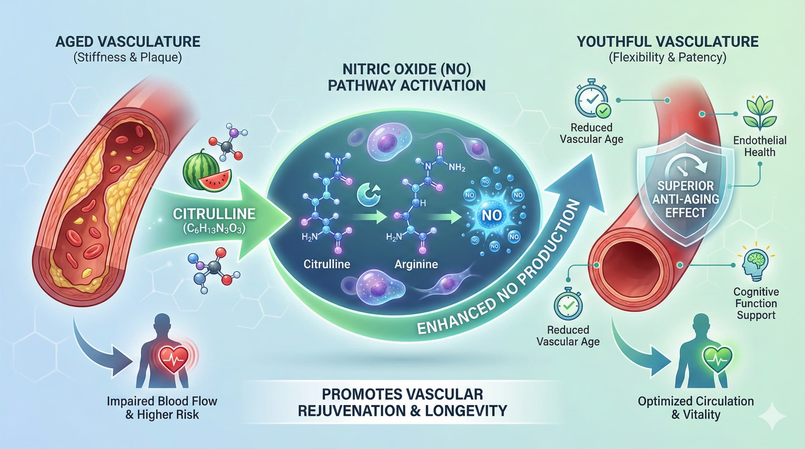
For decades, the “Arginine Paradox” has frustrated longevity researchers: L-arginine is the fuel for Nitric Oxide (NO)—the molecule that keeps arteries young—yet supplementing it often fails to stop vascular aging. A foundational study in PLOS ONE (Tsuboi et al.), now bolstered by 2025 clinical data, confirms why: L-citrulline, not arginine, effectively reverses endothelial senescence.

In diabetic and high-glucose models, arginine supplementation backfires. It triggers Arginase-II, an enzyme that not only diverts fuel away from NO production but actually accelerates cellular aging. Citrulline, acting as a metabolic “Trojan Horse,” bypasses this trap entirely.

The Mechanism: Stealth Delivery

The endothelium (the inner lining of blood vessels) dictates biological age. When stressed by high glucose (diabetes) or age, the endothelium activates senescence pathways (p16-INK4a), stops dividing, and stiffens.

  • Arginine Failure: When you ingest L-arginine, much of it is destroyed by the liver (via arginase). Worse, in aged vessels, extra extracellular arginine upregulates Arginase-II, increasing superoxide production and decoupling the eNOS enzyme.
  • Citrulline Success: L-citrulline bypasses the liver and enters endothelial cells via neutral amino acid transporters. Once inside the cell, it is converted back into arginine locally. This “stealth delivery” restores NO production, suppresses the ROS-generating enzyme NOX2, and—crucially—restores telomerase activity to delay senescence (as detailed in Tsuboi et al. 2018).

The 2025 Evidence Update

While the mechanistic groundwork was laid in 2018, recent data validates the translation to humans:

  • Systemic Anti-Aging: Xie et al. (Science Advances, 2025) recently demonstrated that citrulline reduces senescence markers and DNA damage across multiple tissues, modulating the mTOR-HIF1α axis.
  • Human Microvasculature: Kang & Figueroa (Nutrients, 2025) reported that 4 weeks of citrulline in Type 2 Diabetics significantly improved microvascular oxygenation and muscle function—benefits not seen with arginine alone.
  • Meta-Analysis: A 2025 review in Frontiers in Nutrition (Luo et al. 2025) confirms that citrulline improves flow-mediated dilation (FMD) and reduces arterial stiffness (PWV) in middle-aged cohorts.

Critical Limitations

  • Context is Key: Citrulline shines in diseased, aged, or high-glucose environments (supported by Mori et al. 2021). In perfectly healthy, young individuals with low oxidative stress, the boost in endothelial function is statistically smaller.
  • No Direct “Clock” Data: We lack data on whether citrulline reverses epigenetic age (GrimAge/DunedinPACE) in human endothelial cells specifically.

Actionable Protocol (N=1)

The Strategy: Replace Arginine with Citrulline to lower vascular resistance and support mitochondrial perfusion.

  • Dose: 3 g L-Citrulline (or 6 g Citrulline Malate 2:1), taken 2× daily.
    • Note: Split dosing is superior due to the half-life of NO metabolites.
  • The “Vascular Stack”:
    • + Nitrates (Beetroot): Targets the salivary NO pathway, covering a different mechanism than citrulline.
    • + Glutathione/NAC: To protect the newly generated NO from oxidative degradation.
  • Metrics to Watch:
    • Blood Pressure: Look for a 5–10 point drop in systolic pressure (Maharaj et al. 2022).
    • Endothelial Function: If available, track Flow-Mediated Dilation (FMD) or Reactive Hyperemia Index (EndoPAT).
    • Subjective: Improved peripheral warmth (hands/feet) and exercise tolerance.

Cost-Effectiveness: High. At roughly $0.30 per day, L-citrulline offers a mechanistic ROI superior to expensive NAD+ precursors for vascular-specific aging.

Disclaimer: All these posts are generated with the help of AI systems, and there could be mistakes. Validate with good medical sources before taking any course of action.

RapAdmin

Rapamycin is the flagship mTOR inhibitor in aging research, with systemic benefits reported in multiple organs. This new study asks a narrower question: can rapamycin help repair an overused Achilles tendon? In a rat model, researchers injected collagenase into the Achilles to mimic tendinopathy, then treated animals with a daily intraperitoneal injection of rapamycin (5 mg/kg). Tendons were followed for 2–8 weeks with histology, immunostaining, Western blotting of the mTOR axis, and 8-week biomechanical testing.

What they found

Collagenase produced a classic degenerative tendon phenotype: disrupted collagen, round-cell infiltration, neovascularization, and progressive chondrometaplasia marked by Col-2 and SOX-9 expression. Rapamycin sharply reduced cell density, improved histologic scores, and almost abolished Col-2/SOX-9 upregulation at 4–8 weeks, indicating suppression of ectopic cartilage formation. Western blot showed that collagenase strongly activated AKT–mTOR–p70/S6K signaling, while rapamycin reversed this phosphorylation, confirming on-target mTOR inhibition and a likely shift toward higher autophagy and lower anabolic drive in the injured tendon.

Mechanically, the picture is less encouraging. Rapamycin normalized tendon diameter but did not restore maximum load, tensile stress, or elastic modulus versus collagenase alone; both injured groups remained mechanically weaker than controls at 8 weeks. The authors argue that while mTOR suppression limits pathological chondrogenesis and may optimize collagen architecture, the autophagy–collagen metabolism balance and needle/collagenase damage together constrain strength recovery.

Mechanistic and longevity relevance

For longevity biohackers, this is organ-specific aging in microcosm. Tendons fail with age partly through aberrant matrix remodeling and maladaptive cell fate (tenocytes drifting toward chondrocyte-like states). By dialing down mTOR, rapamycin appears to steer tendon repair away from cartilage-like scarring and toward more tendon-like tissue. Autophagy is implicated but not directly measured; AMPK, cGAS–STING, mitochondrial and vascular endpoints are absent, so any claims about systemic “inflammaging” modulation remain speculative.

What is genuinely novel here is the clear decoupling between histologic “youthfulness” and mechanical function in an Achilles model under explicit mTOR blockade, plus a candid discussion that older animals might show stronger rapamycin responses but were not tested.


Critical Limitations and Translational Uncertainty

  • Model mismatch: acute collagenase + needle damage, not chronic overload or age-related degeneration.
  • Young animals only; rapamycin as an “anti-aging” drug was not tested in aged tendons.
  • High-dose daily IP regimen, unlike intermittent low-dose human longevity protocols.
  • Biomechanics not rescued: the single most relevant endpoint for function remains unimproved.
  • No direct measures of autophagy flux, mitochondrial quality, cGAS–STING, or vascular remodeling; mechanism is inferred from mTOR phosphorylation rather than fully mapped.
  • Follow-up only to 8 weeks; longer-term remodeling unknown.

Further needed: aged-animal models, chronic overload designs, varied rapamycin schedules (including intermittent), direct autophagy/mitochondrial and inflammatory readouts, and human data where tendon structure and function are pre-specified outcomes in rapalog trials.


Full paper (open access) here: Rapamycin-mediated inhibition of the mTOR pathway promotes tendon healing in a collagenase-induced achilles tendinopathy

Disclaimer: All these posts are generated with the help of AI systems, and there could be mistakes. Validate with good medical sources before taking any course of action. This is provided for information purposes only, no medical advice is given here

RapAdmin

A paradigm shift in longevity research is underway, driven by the realization that anti-aging interventions do not confer uniform benefit across the lifespan. Researchers utilizing data from the NIA Interventions Testing Program (ITP) introduced the Temporal Efficacy Profiler (TEP), a novel nonparametric statistical method that accurately estimates time-varying hazard ratios, thereby overcoming the critical limitation of the proportional hazards assumption in the standard log-rank test.

Mechanistic and Temporal Efficacy

The TEP analysis applied to 42 compounds tested in genetically heterogeneous mice is genuinely novel in revealing that efficacy is highly age-dependent, identifying 22 life-extending agents—more than double the prior count—but also uncovering 15 agents that increased mortality risk during specific windows. Critically, most compounds were effective only within restricted age ranges, with only eight agents demonstrating a robust reduction in mortality hazard during the senescence phase (after 90% mortality).

This temporal distinction offers crucial mechanistic insight. Early-life efficacy likely correlates with optimization of growth pathways (e.g., sub-inhibitory mTOR modulation) or metabolic set-points. Interventions like Rapamycin (an mTORC1 inhibitor) or Acarbose (a glucose regulator) showing strong midlife benefits suggest this window is optimal for driving preventative maintenance—specifically the clearance of dysfunctional organelles via autophagy (activated by AMPK) and restoration of mitochondrial quality control, mitigating the onset of widespread metabolic decline. Conversely, the scarcity of late-life effective compounds indicates the difficulty of reversing profound systemic damage, implying that senescence-phase interventions must target persistent burdens like cGAS-STING-driven inflammation, irreversible tissue fibrosis, and vascular stiffening, rather than simply metabolic flux.

Critical Limitations and Cost-Effectiveness

Despite its power, TEP is a statistical model and does not define the underlying molecular mechanisms; its findings are correlational. Data translation is fraught with uncertainty, as the results are based on mouse models (albeit heterogeneous ones). The study relies on all-cause mortality, masking potential organ-specific aging effects (e.g., cardiovascular benefit vs. cognitive impairment). Further data is urgently needed, specifically the integration of TEP outputs with multi-omic data to confirm that the observed temporal efficacy windows correspond precisely to pathway activity (e.g., confirmed AMPK/autophagy upregulation).

For the longevity biohacker, TEP fundamentally shifts safety and cost-effectiveness. By defining the precise window of maximum marginal benefit (and avoiding compounds during periods of life when they are potentially harmful), interventions previously deemed low ROI due to continuous, lifelong administration become highly efficient when pulsed or timed strategically.


Actionable Insights for the Research-Literate Biohacker

  • Biomarkers to Track: Incorporate longitudinal tracking of biological age clocks (e.g., DNAm PhenoAge, GrimAge) to personalize the definition of “midlife” and “senescence” onset, tailoring intervention timing to individual biological, rather than chronological, age.
  • Stacking Hypotheses: Design compound stacks based on non-overlapping temporal efficacy (e.g., using a midlife metabolic agent to prevent damage, followed by a senescence-phase agent to manage established pathology).
  • Dose-Timing Ideas:Explorar pulsatile or cyclic dosing protocols that are restricted to the predicted efficacy window (e.g., a “metabolic reset” cycle started once the PhenoAge clock accelerates, rather than continuous dosing).
  • Implications: Prioritize compounds that demonstrate late-life mortality reduction, as these hold the greatest promise for extending true healthspan and mitigating the primary causes of vascular and cognitive failure.

Specific Compound Timing Data from the Research

Based on the data extracted from the NIA Interventions Testing Program (ITP) re-analysis using the Temporal Efficacy Profiler (TEP), I have reconstructed the specific timing and sex-dependent profiles for the most actionable compounds.

The analysis below isolates the “Elite Eight” (compounds that work in the senescence phase), the “Hidden Hazards” (compounds that turn toxic late in life), and the Sex-Specific agents. And then we have a full list of all 42 compounds with detailed sex and temporal considerations for each compound.

Note: Full details on all the compounds are in the next post in this thread.

Sex-Specific “Non-Responders”

The TEP analysis clarified that “average” data hides the fact that many drugs simply do not work for one sex.

  • Protandim (Nrf2 Activator): Extended median lifespan in Males only. No benefit for Females.
  • Nordihydroguaiaretic Acid (NDGA): Extended median lifespan in Males only. No benefit for Females.
  • Fish Oil (EPA/DHA): TEP analysis confirmed NO significant lifespan extension for either sex in the ITP cohorts tested (though healthspan/cardiovascular specific benefits might exist, they did not translate to all-cause mortality reduction).
  • Curcumin: TEP analysis confirmed NO significant lifespan extension for either sex. (Poor bioavailability is the suspected culprit; TEP confirms standard supplementation is likely useless for longevity).

Summary of “No Effect” / Failed Compounds

According to the ITP and TEP re-analysis, the following popular biohacking compounds showed no statistically significant mortality benefit in the tested doses/formulations:

  • Fisetin (at the continuous dose tested; intermittent high-dose “hit and run” senolytic protocols were not tested in this specific continuous-dosing dataset).
  • Methylene Blue (Showed a small, marginal effect in females in one site, but TEP classifies it as weak/inconsistent compared to the Elite Eight).
  • Simvastatin (No lifespan benefit).
  • Medium-Chain Triglycerides (MCT Oil) (No lifespan benefit).

Biohacker “Cheat Sheet” for Protocol Design

  1. For Males: The “Golden Stack” supported by TEP is Rapamycin + Acarbose + 17-alpha-Estradiol. (Note: 17-alpha-Estradiol is non-feminizing but requires a prescription/compounding). Canagliflozin is a viable late-life addition.

  2. For Females: The protocol is much narrower. Rapamycin is the singular powerhouse. Metformin can be added if stacked with Rapamycin. Avoid Canagliflozin and Green Tea Extract in later years.

  3. The “Fibrosis” Variable: The identification of Halofuginone and Epicatechin as late-life specialists suggests that fighting tissue fibrosis (stiffening of organs) is a distinct, valid target for the senescence phase, separate from metabolic (mTOR/IGF-1) modulation.

Full Open Access Paper: Deciphering the timing and impact of life-extending interventions: temporal efficacy profiler distinguishes early, midlife, and senescence phase efficacies

Disclaimer: All these posts are generated with the help of AI systems, and there could be mistakes. Validate with good medical sources before taking any course of action.

RapAdmin

Longevity research has long treated the kidney as a passive victim of aging, merely a filter that clogs over time. A comprehensive new review challenges this view, positing that the kidney acts as a central “pacemaker” for systemic aging. The mechanism is a vicious feedback loop between cellular senescence and “inflammaging”—chronic, sterile inflammation that accelerates decline across all organ systems.

The core pathology identified is the “Senescence-Associated Secretory Phenotype” (SASP). As renal cells age, they accumulate mitochondrial dysfunction, leading to cytosolic DNA leakage that likely activates the cGAS-STING pathway and NF-κB signaling. These cells do not merely stop working; they become “zombie” cells, pumping out pro-inflammatory cytokines like IL-6 and TNF-α. This local inflammation triggers fibrosis (scarring) and, crucially, downregulates the production of α-Klotho.

Klotho is the study’s linchpin. Produced primarily in the kidney, it is a potent circulating anti-aging protein that regulates phosphate metabolism and protects vascular health. The authors argue that the senescence-induced suppression of Klotho turns a local renal problem into a systemic catastrophe, accelerating vascular calcification, cognitive decline, and sarcopenia. This suggests that interventions preserving renal Klotho output could yield disproportionate benefits for whole-body healthspan.

The review evaluates two primary pharmacological strategies: senolytics (drugs like Dasatinib and Quercetin that induce apoptosis in senescent cells) and senomorphics (compounds like Rapamycin that suppress SASP via mTOR inhibition without killing the cell). The data indicates that clearing renal senescent cells does not just restore kidney function; it breaks the systemic inflammatory loop, restoring Klotho levels and reducing biological age markers in distal tissues.

AI Analysis (from Gemini):

This paper, titled “From Senescent Cells to Systemic Inflammation: The Role of Inflammaging in Age-Related Diseases and Kidney Dysfunction”, explores the biological “vicious cycle” between cellular senescence (aging cells that refuse to die) and inflammaging (chronic, low-grade inflammation). It specifically highlights the kidneys not just as victims of aging, but as central players that can accelerate whole-body aging when they malfunction.


Detailed Breakdown

1. The Core Mechanism: The “Inflammaging” Loop

  • Inflammaging: A chronic, sterile (non-bacterial) inflammation that increases with age. It is predicted by markers like IL-6, TNF-α, and CRP.
  • Immunosenescence: The immune system ages too. It becomes overactive (autoimmunity/inflammation) but less effective at fighting actual threats (infections/cancer).
  • The SASP Factor: Senescent cells stop dividing but remain metabolically active, churning out a “SASP” (Senescence-Associated Secretory Phenotype) cocktail of pro-inflammatory cytokines that turn neighboring healthy cells into senescent ones.

2. The Kidney as an Aging Accelerator

The paper posits the kidney is a “sentinel” of aging.

  • Fibrosis: Chronic inflammation causes renal tissue to scar (fibrosis), reducing function.
  • Klotho Deficiency: The kidney is the main producer of α-Klotho, a powerful anti-aging protein. Inflammation suppresses Klotho production. Lower Klotho levels are directly linked to faster aging, vascular calcification, and cognitive decline.

3. Therapeutic Strategies

The paper reviews two main classes of interventions:

  • Senolytics: Drugs/compounds that selectively kill senescent cells.
  • Senomorphics: Drugs/compounds that suppress the toxic SASP signals without killing the cell.

Actionable Insights for Longevity Biohackers

Based on the interventions reviewed in the paper, here is a list of actionable insights.
> Note: “Preclinical” means the strongest evidence is currently in mice/cells. “Clinical” implies human trial data exists.

1. The Senolytics (The “Clean-Up” Crew)

These compounds aim to periodically purge “zombie” cells to lower the total inflammatory burden.

  • Fisetin (The Accessible Bio-Flavonoid)
    • Paper Finding: Reduces SASP gene expression and preserves kidney histology; protects mitochondria.
    • Biohacker Protocol: Fisetin is widely available as a supplement. Biohackers often use “hit-and-run” pulsing protocols (e.g., high dose for 2-3 days, then off for a month) rather than daily low dosing, mimicking the intermittent “clearing” seen in studies.
    • Evidence Level: Preclinical (mixed), Clinical trials ongoing.
  • Quercetin + Dasatinib (The “D+Q” Combo)
    • Paper Finding: One of the most potent combinations. It targets anti-apoptotic pathways to force senescent cells to die. Shown to reduce SASP, improve renal markers, and—crucially—increase α-Klotho expression.
    • Biohacker Protocol: Quercetin is a common supplement (best taken with bromelain or in phytosome form for absorption). Dasatinib is a prescription leukemia drug and carries significant risks/side effects. Advanced biohackers often focus on Quercetin alone or combine it with other natural senolytics if they cannot access or do not want to risk Dasatinib.
    • Evidence Level: Preclinical + Clinical.
  • Caution on Dasatinib: Dasatinib and Quercetin as Senolytic May Cause Brain Damage

2. The Senomorphics (The “Quieting” Agents)

These are typically taken more regularly to suppress the inflammatory noise (SASP) from aging cells.

  • Rapamycin (mTOR Inhibition)
    • Paper Finding: Suppresses SASP and fibrosis by inhibiting the mTOR pathway.
    • Biohacker Protocol: Currently the “gold standard” prescription longevity drug in the biohacking community. Usually taken once weekly (pulsed) to minimize immune suppression while maximizing mTOR inhibition.
    • Evidence Level: Strong Preclinical; growing human safety data.

3. Kidney-Specific Protection (The “Klotho” Strategy)

Since the kidney produces the anti-aging protein Klotho, protecting kidney function is a longevity strategy in itself.

  • Monitor Cystatin C: Don’t just rely on Creatinine/eGFR blood tests. Cystatin C is a more sensitive marker of kidney function that this paper’s context suggests would be valuable for early detection of dysfunction.
  • Prioritize Mitochondrial Health: The paper links mitochondrial dysfunction directly to kidney inflammaging. Interventions like CoQ10, PQQ, and Urolithin A (mitophagy inducers) are logically consistent additions to support the high-energy demands of kidney cells.

4. Emerging/Experimental Targets

  • FOXO4-DRI: A peptide that disrupts the interaction between FOXO4 and p53, causing senescent cells to commit suicide. (Mentioned as effective in enhancing renal function in preclinical models, but currently difficult to source reliably for humans).
  • Glutaminase Inhibitors: Targeting the metabolism of senescent cells (which are addicted to glutamine). This suggests that cycling glutamine or fasting might mimic some of these benefits, though the paper focuses on pharmaceutical inhibitors.

Disclaimer: All these posts are generated with the help of AI systems, and there could be mistakes. Validate with good medical sources before taking any course of action.

RapAdmin

Yet another positive study, in a vast ocean of favorable rapamycin research (in animals).

A new long-term study of intermittent, low-dose Rapamycin (RAPA) in mice demonstrates preservation of tissue function, attenuation of age-associated pathology, and delay of age-related frailty metrics — using a pulsed, low-dose administration.

In this work, mice received RAPA at doses roughly equivalent to a low human-equivalent dose, administered intermittently over their lifespan. Compared with controls, treated mice showed improved markers of organ integrity, reduced degenerative changes, and slower onset of dysfunction across multiple systems — suggesting “quality of life” gains rather than extreme lifespan extension.

Mechanistically, the outcomes likely stem from modulation of the mTOR pathway, periodic activation of autophagy, enhanced mitochondrial maintenance, and possibly improved vascular homeostasis — while avoiding chronic suppression that might provoke compensatory stress or immune dysregulation. The intermittent schedule may also reduce negative feedback on upstream regulators like AMPK, preserving metabolic flexibility. Though the authors do not report direct measures of vascular or cGAS-STING inflammation, the pattern of reduced tissue pathology is consistent with lower chronic inflammatory/ senescence-associated signaling.

What’s slightly novel is the demonstration that low-dose, intermittent RAPA — rather than higher dosing — can yield sustained tissue benefits over a full lifespan, suggesting another intervention framework relevant to human longevity use. Prior studies with rapamycin have tended to use higher doses to maximize longevity benefits; this study reframes RAPA as a potential low-dose, chronic “maintenance” drug.

Possible Actionable Hypotheses for n=1 Biohackers

  • Track biomarkers: periodic measurement (e.g. quarterly) of CRP / IL-6 (inflammation), ALT/AST (liver), eGFR + cystatin-C (kidney), HbA1c + fasting glucose (metabolic), and lipid panel + pulse-wave velocity or ankle-brachial index (vascular) to assess tissue-level burden.
  • Consider an intermittent low-dose rapamycin protocol (e.g., 2–3 mg once weekly or biweekly, depending on weight/age, analogous to mouse equivalence), rather than daily or high-dose — to maximize safety/benefit ratio.
  • Evaluate stacking with AMPK activators (e.g., low-dose Metformin or exercise/fasting mimetics) to preserve metabolic flexibility while using RAPA; hypothesize synergy via alternating autophagy and metabolic stress pathways.
  • Cycle rapamycin seasonally or quarterly to monitor longitudinal effects, avoid receptor down-regulation, and better detect on-off phase functional changes.
  • Monitor for trade-offs: track immune function (CBC with differential, blood glucose levels, vaccine response if feasible) to avoid subtle immunosuppression or impaired wound/repair function.

From a cost-effectiveness standpoint, rapamycin at low intermittent dosing is relatively inexpensive compared with many longevity interventions (stem-cell therapies, gene therapy, frequent diagnostics), offering potentially high marginal benefit per dollar — especially if maintained long-term with minimal side-effects.

Critical Limitations & Translational Uncertainty

  • The data derive from mice; human biology — especially dose scaling, immune system complexity, and long-term tolerability — may differ.
  • The study lacks direct measures of autophagy flux, mitochondrial function, vascular function, senescence markers, or cGAS-STING/inflammatory signaling — leaving mechanistic inferences speculative rather than demonstrated.
  • Effect-size uncertainty remains: while organ-level preservation is shown, impact on maximum lifespan, compression of morbidity, or late-life resilience remains unclear.

To fully translate into human use, trials need: longitudinal biomarker tracking under low-dose intermittent rapamycin; tissue-level readouts (e.g. via imaging or functional assays) rather than only surrogate serum markers; and eventual controlled human studies assessing frailty, morbidity compression, and safety over years.


Source Paper (Open access): Effects of Long-term low-dose intermittent rapamycin administration on glucose metabolism and immune system of SAMP8 and SAMR1 mice

Note: There seems to be a typo in the paper: it says dosing of 0.78 ug/kg, but I suspect its mg/kg.

In-depth CGPT analysis of paper: Journal article evaluation

Relacionado:

RapAdmin

A new Aging Cell study delivers one of the most granular views to date of how long-term caloric restriction (CR) reshapes the aging primate brain. Using single-nucleus RNA sequencing on postmortem tissue from elderly rhesus monkeys, the researchers uncovered profound shifts in the biology of two cell populations essential for brain maintenance: oligodendrocytes, which produce myelin, and microglia, the brain’s resident immune cells.

The results point to a coordinated transcriptional program induced by decades of reduced calorie intake—approximately 30% below ad libitum feeding—one that appears to bolster myelin integrity and reduce inflammation-linked degeneration. Oligodendrocytes from the CR animals showed enhanced expression of genes supporting glycolysis, fatty-acid biosynthesis, and myelin production. Some oligodendrocyte subtypes also displayed strengthened adhesion to axons, suggesting tighter structural coupling and potentially more resilient white-matter tracts. Meanwhile, microglia in CR brains exhibited increased metabolic activity and a marked reduction in gene signatures associated with myelin debris accumulation. This pattern suggests either more efficient debris clearance or less upstream myelin damage — both consistent with preserved neural circuitry during aging.

These cellular findings align with broader evidence that CR slows systemic aging, lowers inflammation, and maintains tissue homeostasis. They also highlight an interesting mechanistic convergence with rapamycin, the best-validated pharmacologic longevity intervention. Both CR and rapamycin modulate nutrient-sensing pathways—particularly mTOR—leading to enhanced cellular stress resistance, improved proteostasis, and shifts toward metabolic efficiency. The primate data hint that CR may exert more targeted effects on glial cell metabolism and myelination than currently demonstrated for rapamycin, whereas rapamycin more consistently boosts autophagy, reduces senescent signatures, and alters immune-cell composition across multiple tissues. The two approaches could be complementary rather than redundant: CR preserving structural brain integrity, rapamycin improving intracellular cleanup and repair.

For longevity-oriented individuals, the translational signals are practical. Sustained, nutrient-adequate caloric restriction may help maintain white-matter quality and glial function with aging. Intermittent CR or CR-mimetic strategies (fasting-mimicking diets, protein restriction windows, or pharmacologic mimetics) might reproduce portions of these benefits without the burden of lifelong restriction. The overlap with rapamycin suggests that dietary energy restriction and mTOR inhibition may reinforce each other’s effects on brain aging, although no human data yet address combined interventions.

The study’s limitations are substantial: small sample size, species differences, absence of cognitive or imaging endpoints, and reliance on transcriptional signatures rather than functional measures. Lifelong CR is also impractical for most humans and may carry risks if poorly designed. Nonetheless, this work provides rare molecular evidence—directly in primate brain tissue—that caloric restriction remodels aging at the level of specific glial cell programs, strengthening the argument that nutrient-sensing pathways remain central leverage points for extending brain healthspan.

Open Access Paper: Calorie Restriction Attenuates Transcriptional Aging Signatures in White Matter Oligodendrocytes and Immune Cells of the Monkey Brain (Aging Cell)

RapAdmin

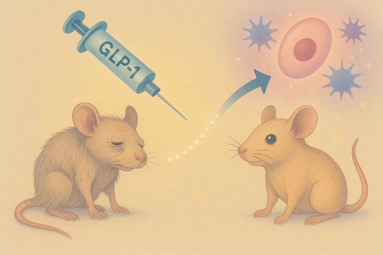
A new Cell Metabolism study reports that GLP-1 receptor agonists—currently known for weight-loss and metabolic control—produce broad, body-wide anti-aging effects in mice independent of weight loss. The work arrives just as the more effective new oral GLP-1 pills are slated for release next year, significantly expanding the practical reach of this drug class and elevating their relevance to the longevity community.

In the study, middle-aged and older male mice received long-term GLP-1RA treatment at doses too low to alter appetite or body weight. This design was deliberate: the goal was to isolate the drug’s intrinsic aging biology effects. After several weeks, researchers conducted deep multi-omic profiling across major organs. The results were unambiguous—tissues ranging from the liver and muscle to the colon, adipose depots, blood, and even regions of the brain shifted toward a younger molecular state. Gene-expression programs normalized, chronic inflammation signatures diminished, metabolic pathways rebalanced, and epigenetic patterns moved in a youth-associated direction. Functional testing echoed these findings: treated older mice exhibited stronger grip and superior motor coordination.

One of the most consequential observations was the overlap between GLP-1–induced rejuvenation patterns and those produced by rapamycin, the canonical mTORC1 inhibitor that has extended lifespan in every mammalian study completed to date. mTORC1 acts as a nutrient-sensing growth switch; when chronically activated, it suppresses autophagy, accelerates protein damage, distorts cellular metabolism, and drives hallmark aging pathology. Rapamycin works by inhibiting this overactive pathway.

GLP-1R agonists engage aging biology differently. Rather than acting directly on nutrient sensing, they recalibrate metabolic and endocrine communication through the hypothalamus, producing downstream effects—enhanced proteostasis, reduced inflammation, metabolic normalization—that converge with the benefits of mTOR inhibition. In practical terms: GLP-1s may achieve a portion of rapamycin’s biological “reset,” but through a neuroendocrine route that could carry fewer immunosuppressive consequences.

For longevity-focused individuals, several actionable insights emerge:

1. GLP-1s may have geroprotective value even at doses below those used for weight loss.

Future human trials may explore “metabolically quiet” doses that avoid appetite suppression while targeting systemic aging pathways.

2. Combining GLP-1s with lifestyle interventions likely produces synergistic benefits.

The molecular changes observed align with improved metabolic flexibility—an effect amplified by resistance training, fasting-mimicking diets, and reduced ultra-processed food intake.

3. With new oral GLP-1 pills arriving soon, adherence, affordability, and long-term deployment will improve dramatically, making them viable candidates in structured, physician-supervised longevity protocols.

4. GLP-1s may serve as an option for individuals unable to tolerate rapamycin due to side-effects, or for those seeking partial mTOR-like benefits without immunosuppression.

Still, major limitations remain. The study used only male mice, leaving female biology unexplored. Cognitive improvements were limited, underscoring that brain aging may require additional modalities (exercise, sleep optimization, neurotrophic support, rapamycin, or senolytics). Most importantly, lifespan extension was not measured—so rejuvenation signatures, while encouraging, require validation in long-term survival and disease-onset studies. And chronic GLP-1 use in healthy humans remains insufficiently studied.

Even with these caveats, the findings suggest a paradigm shift: GLP-1 medications—soon available as both injections and moderately expensive pills—may represent one of the first widely accessible pharmacological tools capable of modulating core mechanisms of aging.

Source Research Paper (Open access): Body-wide multi-omic counteraction of aging with GLP-1R agonism

CGPT In-depth analysis of paper: https://chatgpt.com/share/6929ef82-baa8-8008-93f1-04d27c98a9e5

Disclaimer: All these posts are generated with the help of AI systems, and there could be mistakes. Validate with good medical sources before taking any course of action.